Comparative Study
. 2009 Jul 8;29(27):8805-15.
doi: 10.1523/JNEUROSCI.6159-08.2009.

Deletion of the alpha 7 nicotinic acetylcholine receptor gene improves cognitive deficits and synaptic pathology in a mouse model of Alzheimer's disease

Affiliations
Comparative Study

Deletion of the alpha 7 nicotinic acetylcholine receptor gene improves cognitive deficits and synaptic pathology in a mouse model of Alzheimer's disease

Gustavo Dziewczapolski et al. J Neurosci. .

Abstract

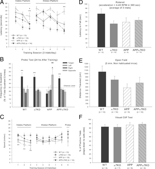
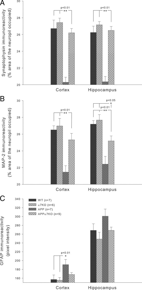
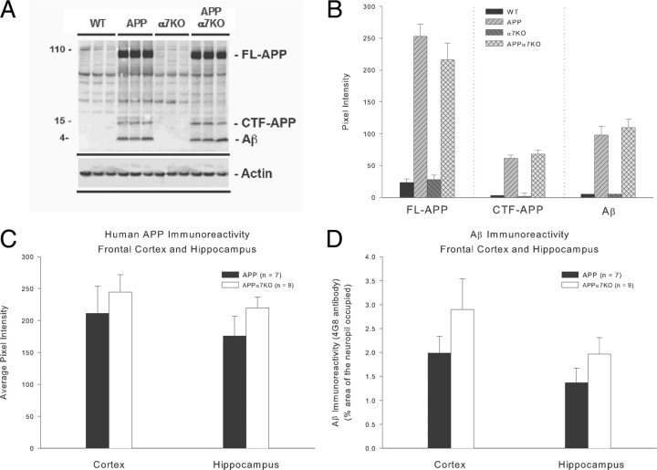
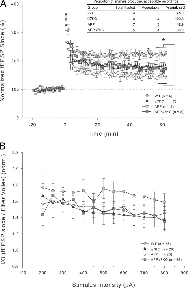
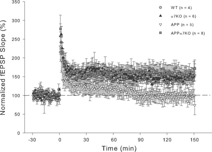
It has been recently shown that the Alzheimer's disease (AD) pathogenic peptide amyloid beta(1-42) (Abeta(1-42)) binds to the alpha7 nicotinic acetylcholine receptor (alpha7nAChR) with high affinity and the alpha7nAChR and Abeta(1-42) are both found colocalized in neuritic plaques of human brains with AD. Moreover, the intraneuronal accumulation of Abeta(1-42) was shown to be facilitated by its high-affinity binding to the alpha7nAChR, and alpha7nAChR activation mediates Abeta-induced tau protein phosphorylation. To test the hypothesis that alpha7nAChRs are involved in AD pathogenesis, we used a transgenic mouse model of AD overexpressing a mutated form of the human amyloid precursor protein (APP) and lacking the alpha7nAChR gene (APPalpha7KO). We have shown that, despite the presence of high amounts of APP and amyloid deposits, deleting the alpha7nAChR subunit in the mouse model of AD leads to a protection from the dysfunction in synaptic integrity (pathology and plasticity) and learning and memory behavior. Specifically, APPalpha7KO mice express APP and Abeta at levels similar to APP mice, and yet they were able to solve a cognitive challenge such as the Morris water maze test significantly better than APP, with performances comparable to control groups. Moreover, deleting the alpha7nAChR subunit protected the brain from loss of the synaptic markers synaptophysin and MAP2, reduced the gliosis, and preserved the capacity to elicit long-term potentiation otherwise deficient in APP mice. These results are consistent with the hypothesis that the alpha7nAChR plays a role in AD and suggest that interrupting alpha7nAChR function could be beneficial in the treatment of AD.


Figures

Figure 1.
Figure 1.
Deletion of the α7nAChR prevents learning and memory deficits observed in APP-overexpressing animals. Water maze performance was evaluated in mice at 13–16 months of age. Mice were tested for a total of 9 d. A, From days 1 to 3, visible platform training was performed to acclimatize the animals to the test conditions and from days 4 to 8; the animals were trained with a hidden platform to test spatial learning abilities. We measured latency to reach the platform. APP mice showed significant spatial learning impairments as a lack of improvement in five consecutive hidden platform sessions (p > 0.05), compared with APP mice lacking functional α7nAChR at levels similar to controls, WT and α7KO mice. For simplification, significant differences between groups are only shown for the last day of spatial learning (**p < 0.01 vs all 3 other groups). Note also that although APP mice significantly improved their performance in the visible platform version of the Morris water maze test (p < 0.05, day 3 vs day 1), they needed more time to reach the platform than all the other three genotypes (**p < 0.01 vs α7KO and *p < 0.05 vs WT or APPα7KO in the last session day). B, On day 9, memory was evaluated with a probe test as the frequency the target quadrant was visited in relation to the other three quadrants. Deletion of the α7nAChR rescued the memory deficit observed in APP-overexpressing animals and reached values similar to controls, WT and α7KO, as shown by a significant increase in frequency to enter the target quadrant (**p < 0.01 vs all 3 other quadrants). Note the inverse frequency in quadrant preference observed in APP mice (##p < 0.01 vs target quadrant and #p < 0.05 vs left and right quadrants). C–F, Motor (C, swim speed; D, rotarod; E, open field) and visual (F, visual cliff test) abilities are not significantly affected in APP mice, suggesting that performance deficits in all aspects of the Morris water maze test are consequence of affected learning and memory mechanisms (see Results for details on each of these four evaluations, especially the complex swim speed analysis). Differences among means were assessed by two-factor (training session and genotype) ANOVA with repeated measures for the training block factor or one-factor (genotype or quadrant) ANOVA followed by post hoc multiple-comparisons Fisher's test. Mean values ± SEM are shown.
Figure 2.
Figure 2.
Deletion of the α7nAChR ameliorates the neuropathology observed in APP-overexpressing animals. Immunohistochemistry for the synaptic terminal marker synaptophysin, the dendritic marker MAP2, and the astrogliosis marker GFAP was evaluated in the frontal cortex and hippocampus of 19- to 22-month-old mice. A, A significant reduction in synaptophysin was observed in both cortex and hippocampus of APP mice compared with APPα7KO mice (p < 0.01). B, MAP2 was also markedly reduced in APP mice in both cortex and hippocampus compared with the normal or nearly normal levels in the cortex (p < 0.01) and hippocampus (p < 0.01 vs APP but also p < 0.05 vs controls, WT and α7KO) of APPα7KO mice, respectively. C, We also measured astrogliosis in frontal cortex and hippocampus using GFAP immunoreactivity as a marker. Immunoreactivity levels were significantly elevated in frontal cortex in APP mice (p < 0.01 vs controls, WT or α7KO). In contrast, GFAP immunoreactivity levels were only partially increased in the APPα7KO mice, not reaching statistical significance compared with WT or α7KO controls or APP mice (p > 0.05). In the hippocampus, APPα7KO mice showed GFAP immunoreactivity values similar to the control groups, while GFAP immunoreactivity levels in the APP group tended (p = 0.063) to be higher. Differences among means were assessed by one-way (genotype) ANOVA followed by post hoc multiple-comparisons Fisher's test. Mean values ± SEM are shown.
Figure 3.
Figure 3.
APP and APPα7KO mice express same levels of Aβ and APP in cortex and hippocampus. We assessed the levels of Aβ and APP expression using Western blot (A, B), immunohistochemistry (C, D), and ELISA (see supplemental Fig. 5, available at www.jneurosci.org as supplemental material) methods, and the results revealed no differences between APP and APPα7KO mice (p > 0.05). In A and B, results are shown for dissected cortex plus hippocampus. Mean values ± SEM are shown.
Figure 4.
Figure 4.
Deletion of α7nAChR prevents LTP deficits observed in APP-overexpressing mice at the Schaffer collateral to CA1 synapse. A, After a 20 min baseline, LTP was induced by four tetani delivered 5 min apart, each at 100 Hz for 1 s. fEPSPs were monitored for 60 min after tetanus. LTP in APP animals was significantly lower than in the control WT group (*p < 0.05). In contrast to APP mice, LTP was normal (no significant differences vs WT) in APPα7KO and α7KO mice groups. The magnitude of this effect may be an underestimate since the data include only those animals in which we could obtain acceptable recordings, and a strikingly high proportion of APP mice did not produce acceptable recordings (see table inset). Differences among means were assessed by one-way (genotype) ANOVA followed by post hoc Dunn's (Bonferroni) correction. B, Basal synaptic transmission was also recorded at the same synapse in a different set of age-matched mice, but no significant differences in the I/O slopes (fEPSP vs fiber volley) between all the four different genotypes were found. Mean values ± SEM are shown.
Figure 5.
Figure 5.
Deletion of α7nAChR in APP-overexpressing mice elicits a normal sustained late-phase LTP compared with the decay observed in APP-overexpressing littermates at the Schaffer collateral to CA1 synapse. After a 30 min baseline, LTP was induced by four tetani delivered 5 min apart, each at 100 Hz for 1 s. fEPSPs were monitored for 150 min after tetanus. LTP in APP animals was short lived during an early phase and rapidly decayed to baseline levels after 1 h. In contrast to APP mice, early and late LTP are normal in APPα7KO mice with no signs of decay and with potency similar to controls, WT and α7KO mice, for the entire 150 min recorded. Mean values ± SEM are shown.

References

    1. Aracava Y, Pereira EF, Maelicke A, Albuquerque EX. Memantine blocks alpha7* nicotinic acetylcholine receptors more potently than N-methyl-d-aspartate receptors in rat hippocampal neurons. J Pharmacol Exp Ther. 2005;312:1195–1205. - PubMed
    1. Armstrong JN, Saganich MJ, Xu N-J, Henkemeyer M, Heinemann SF, Contractor A. B-Ephrin reverse signaling is required for NMDA-independent long-term potentiation of mossy fibers in the hippocampus. J Neurosci. 2006;26:3474–3481. - PMC - PubMed
    1. Banerjee P, Samoriski G, Gupta S. Comments on “Memantine blocks {alpha}7* nicotinic acetylcholine receptors more potently than N-methyl-d-aspartate receptors in rat hippocampal neurons.”. J Pharmacol Exp Ther. 2005;313:928–929. author reply 930–933. - PubMed
    1. Bednar I, Paterson D, Marutle A, Pham TM, Svedberg M, Hellström-Lindahl E, Mousavi M, Court J, Morris C, Perry E, Mohammed A, Zhang X, Nordberg A. Selective nicotinic receptor consequences in APP(SWE) transgenic mice. Mol Cell Neurosci. 2002;20:354–365. - PubMed
    1. Billings LM, Oddo S, Green KN, McGaugh JL, LaFerla FM. Intraneuronal Abeta causes the onset of early Alzheimer's disease-related cognitive deficits in transgenic mice. Neuron. 2005;45:675–688. - PubMed

Publication types

Substances