Comparative Study
. 2009 Jul 8;29(27):8858-70.
doi: 10.1523/JNEUROSCI.1423-09.2009.

Dlg1, Sec8, and Mtmr2 regulate membrane homeostasis in Schwann cell myelination

Affiliations
Comparative Study

Dlg1, Sec8, and Mtmr2 regulate membrane homeostasis in Schwann cell myelination

Annalisa Bolis et al. J Neurosci. .

Abstract

How membrane biosynthesis and homeostasis is achieved in myelinating glia is mostly unknown. We previously reported that loss of myotubularin-related protein 2 (MTMR2) provokes autosomal recessive demyelinating Charcot-Marie-Tooth type 4B1 neuropathy, characterized by excessive redundant myelin, also known as myelin outfoldings. We generated a Mtmr2-null mouse that models the human neuropathy. We also found that, in Schwann cells, Mtmr2 interacts with Discs large 1 (Dlg1), a scaffold involved in polarized trafficking and membrane addition, whose localization in Mtmr2-null nerves is altered. We here report that, in Schwann cells, Dlg1 also interacts with kinesin 13B (kif13B) and Sec8, which are involved in vesicle transport and membrane tethering in polarized cells, respectively. Taking advantage of the Mtmr2-null mouse as a model of impaired membrane formation, we provide here the first evidence for a machinery that titrates membrane formation during myelination. We established Schwann cell/DRG neuron cocultures from Mtmr2-null mice, in which myelin outfoldings were reproduced and almost completely rescued by Mtmr2 replacement. By exploiting this in vitro model, we propose a mechanism whereby kif13B kinesin transports Dlg1 to sites of membrane remodeling where it coordinates a homeostatic control of myelination. The interaction of Dlg1 with the Sec8 exocyst component promotes membrane addition, whereas with Mtmr2, negatively regulates membrane formation. Myelin outfoldings thus arise as a consequence of the loss of negative control on the amount of membrane, which is produced during myelination.


Figures
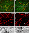
Figure 1.
Figure 1.
Myelin outfoldings are reproduced in Mtmr2-null Schwann cells/DRG neuron cocultures. Control and Mtmr2-null Schwann cell/DRG neuron cocultures were treated for 15 d with ascorbic acid to induce myelination. A, A′ inset, C, After MBP labeling, myelinated fibers had a normal shape in control. B, B′ inset, D, Abnormal shape in Mtmr2-null explants. A, B, Neurofilament (NF) was used to detect axons. E–H, Electron microscopy demonstrated that myelin outfoldings were present in myelin-forming cocultures. The arrowheads in B′, D, and F–H indicate myelin outfoldings. The myelin outfolding phenotype is fully penetrant in vitro, as at least 40 different Mtmr2-null embryos were evaluated in this work. Scale bars: (in A) A, B, 10 μm; A′, B′, C, D, 7 μm; (in H) E–H, 1 μm.
Figure 2.
Figure 2.
Loss of Mtmr2 and Mtmr2/Dlg1 interaction causes myelin outfoldings in vitro. Myelin outfoldings are rescued by MTMR2 replacement using LV transduction. A, Transduction of Mtmr2-null Schwann cell/DRG neuron cocultures with a GFP LV. A′, Inset, MBP-positive fibers with myelin outfoldings are indicated by arrowheads. B, Transduction of Mtmr2-null Schwann cell/DRG neuron cocultures with FLAG-MTMR2 LV rescues myelin outfoldings. DAPI, 4′,6′-Diamidino-2-phenylindole. B′, Inset. C, Electron microscopy on FLAG-MTMR2 LV transduced cocultures showing that myelin compaction is preserved on MTMR2 overexpression. At least 30 myelinated fibers were evaluated and compared with cultures infected with GFP alone. D, Quantification of MBP-positive fibers with myelin outfoldings in not transduced (NT) explants (75.5 ± 2.3; n = 6), transduced with GFP alone (76.5 ± 5.0; n = 4), and with FLAG-MTMR2 (10.7 ± 5.3; n = 9) (p = 8.7 × 10⁻⁶) (from at least 2 different experiments). E, Dlg1 coimmunoprecipitates with FLAG-MTMR2 but not with MTMR2 devoid of the PDZ-binding domain at the C terminus, termed FLAG-MTMR2ΔPDZBD. Input is the lysate. F, Western blot analysis showing similar level of expression of both FLAG-MTMR2 and FLAG-MTMR2ΔPDZBD on LV transduction of Mtmr2-null DRG explants at MOI12.5. G, H, Examples of myelin-forming cocultures transduced with FLAG-MTMR2 or FLAG-MTMR2ΔPDZBD LV at MOI12.5. I, Quantification of MBP-positive fibers with myelin outfoldings on LV transduction of Mtmr2-null explants with FLAG-MTMR2 (17.99 ± 2.20; n = 9) and FLAG-MTMR2ΔPDZBD (29.97 ± 1.76; n = 9) (p = 0.00085); FLAG-MTMR2ΔPDZBD (29.97 ± 1.76; n = 9) and GFP (76.5 ± 5.0; n = 4) (p = 0.00097) (2 different experiments). Error bars indicate SEM. **p < 0.001; ***p <0.0001. Scale bars: (in H) A, B, 20 μm; A′, B′, 14 μm; G, H, 10 μm; (in C) C, 100 nm.
Figure 3.
Figure 3.
Dlg1 interacts with kif13B. A–E, Dlg1/kif13B interaction was further demonstrated by performing pull-down assays on P11 rat nerve lysates using GST-Dlg1/GUK and GST-kif13B/MBS, in which the input is the nerve lysate. C, Western blot analysis confirmed kif13B expression in isolated rat Schwann cells. F, G, In vivo, in transverse rat nerves, kif13B is expressed in the cytosol of both myelin-forming and in non-myelin-forming Schwann cells, colocalizing with the cytosolic marker S100. G, GFAP staining identified non-myelin-forming Schwann cells. H, kif13B is also expressed in axons, although less abundantly. I, In teased-fiber analysis, kif13B expression was detected in internodal cytoplasmic channels and microvilli–paranodes as indicated by arrows (node). J, kif13B was also detected in SLIs. L–O, Immunohistochemistry performed on Mtmr2-null fibers showed normal kif13B localization. The arrows indicate SLIs in M, and, in O, perinuclear staining. K, kif13B expression level in Mtmr2-null nerves is not altered as shown by Western blot analysis. Sciatic nerves from 2- to 3-month-old animals were used. Scale bars, 10 μm.
Figure 4.
Figure 4.
Dlg1 interacts with Sec8. A, Coimmunoprecipitation of Dlg1 and Sec8 from mouse nerves. Input is the lysate; “unb” is the unbound fraction of the lysate after immunoprecipitation. B, Western blot analysis showing Sec8 expression in isolated rat Schwann cells, with MDCK cell lysate as control. C, Western blot analysis confirmed that Sec8 expression level in Mtmr2-null nerve lysate is not altered. D–F, Immunohistochemistry on transverse rat nerve sections revealed a Sec8 punctate staining in the cytoplasm of myelin-forming Schwann cells, S100 positive, but not in non-myelin-forming Schwann cells, GFAP positive, nor in axons. G, H, Immunohistochemistry on rat teased nerves showed Sec8 expression in SLIs (arrowheads) and in the microvilli–paranodes. Caspr is a marker of paranodes in the axon. I, A strong Sec8 perinuclear expression was also detected as shown by colocalization with the Golgi marker Giantin. J, K, Immunohistochemistry on Mtmr2-null mouse nerves, showing that Sec8 expression at microvilli is not altered with respect to control fibers. Sciatic nerves from 2- to 3-month-old animals were used. Scale bars: (in F) D–F, 5 μm; (in K) G–I, 10 μm; J, K, 5 μm.
Figure 5.
Figure 5.
GTS-kif13B/MBS pulls down Dlg1, Sec8, and Mtmr2 in both nerve and isolated Schwann cells. A, Pull-down assay using GST-Mtmr2 on P11 rat nerves. Sec8 and Dlg1 were revealed by Western blot analysis. B, The same experiment was performed from isolated rat Schwann cells lysate to confirm that these interactions likely occur in Schwann cells in the nerve. C, D, Pull-down assays using GST-kif13B/MBS from P11 rat nerve and isolated rat Schwann cell lysates, in which Sec8, Dlg1, and Mtmr2 were detected. Mtmr2-null and wild-type mouse nerve lysates were also used as control to test the specificity of the anti-Mtmr2 antibody used. Two bands of ∼70 and 60 kDa disappear in the Mtmr2-null nerve lysate, suggesting that this antibody recognizes Mtmr2. Input is the lysate, and “unb” is the unbound fraction after pull down.
Figure 6.
Figure 6.
kif13B transports Dlg1 in Schwann cells. A, Dominant-negative constructs of kif13B used in myelin-forming cocultures. B, C, Coimmunoprecipitation in COS7 cells between overexpressed myc-kif13BΔMD and Dlg1 and between overexpressed myc-kif13B/MBS and Dlg1. D, Western blot analysis on lysates from myelin-forming cocultures transduced with myc-kif13BΔMD LV, in which control is the GFP LV. E, myc-kif13BΔMD LV is expressed in almost all cells with high copy number integration. F, G, Dlg1 localization in myelin-forming cocultures appears at microvilli similar to what has been observed in immature nerves in vivo during postnatal development (see also supplemental Fig. 2A–C, available at www.jneurosci.org as supplemental material). H–J, Quantification of Dlg1-positive clusters in myelinated fibers stained with MBP on myc-kif13BΔMD overexpression (myc-kif13BΔMD, 50.74 ± 5.04, n = 6; GFP, 78.40 ± 3.49, n = 7; p = 0.00146; 2 experiments). L, Western blot analysis on lysates from myelin-forming cocultures transduced with myc-kif13B/MBS LV, in which control is GFP LV. K–N, Quantification of MBP-positive fibers in explants overexpressing myc-kif13B/MBS (myc-kif13B/MBS, 46.17 ± 8.50, n = 6; GFP, 100 ± 11.07, n = 4; p = 0.00997). Error bars indicate SEM. *p < 0.05. Scale bars: (in J) I, J, 10 μm; E, 30 μm; F, G, 2 μm; (in N) M, N, 100 μm.
Figure 7.
Figure 7.
Loss of kif13B and Dlg1 in myelin-forming cocultures impairs active myelination. A, kif13B shRNA LV transduction of isolated rat Schwann cells revealed a decrease of kif13B, Dlg1, and Sec8 expression. Three independent experiments were performed. B, kif13B shRNA LV transduction of dissociated Schwann cell/DRG neuron cocultures also revealed a decrease of kif13B, Dlg1, and Sec8 expression. C, Downregulation of Dlg1 expression on Dlg1 shRNA LV transduction of isolated rat Schwann cells. D–F, In dissociated cultures transduced with kif13B shRNA LV, myelination, as the ratio between the number of MBP-positive segments and number of Schwann cell nuclei, was significantly decreased with respect to control shRNA (kif13B shRNA, 34.80 ± 14.73, n = 12; control shRNA, 100 ± 16.60, n = 16; p = 0.00779; 2 experiments). G–I, The amount of MBP-positive segments is also decreased in Schwann cell/DRG neuron cocultures on Dlg1 shRNA transduction (Dlg1 shRNA, 19 ± 10.2, n = 7; control shRNA, 100 ± 25.5, n = 7; p = 0.0107). Error bars indicate SEM. *p < 0.05. Scale bar, 50 μm.
Figure 8.
Figure 8.
Sec8 is involved in myelin formation in Schwann cells. A–D, A significant decrease in myelination was observed in dissociated Schwann cell/DRG neuron cocultures on Sec8 shRNA LV transduction (Sec8 shRNA, 28.01 ± 5.95, n = 6; control shRNA, 100 ± 18.01, n = 10; p = 0.00296). B, The amount of MBP-positive segments was normalized to the Schwann cell number. E–G, Dissociated cocultures were transduced with different amounts of Sec8 shRNA LV. The highest concentration of LV that did not affect myelination was chosen to transduce Mtmr2-null dissociated cocultures. H–J, Myelin outfoldings were significantly rescued also when myelination is not impaired (Sec8 shRNA, 51.79 ± 1.67, n = 6; control shRNA, 64.78 ± 3.22, n = 6; p = 0.00716). Error bars indicate SEM. *p < 0.05. Scale bars: (in G) C, D, F, G, 50 μm; (in J) I, J, 10 μm.
Figure 9.
Figure 9.
Proposed model of kif13B, Dlg1, Mtmr2, and Sec8 interactions at sites of Schwann cell membrane homeostasis. We propose a model by which Dlg1, transported by kif13B, organizes a molecular platform to coordinate the Sec8-mediated function on membrane formation with that of Mtmr2 on membrane remodeling to achieve homeostasis. Mtmr2 might be either transported via kif13B/Dlg1 (1) or clustered at the sites of membrane homeostasis (2).

Similar articles

Cited by

References

    1. Anitei M, Ifrim M, Ewart MA, Cowan AE, Carson JH, Bansal R, Pfeiffer SE. A role for Sec8 in oligodendrocyte morphological differentiation. J Cell Sci. 2006;119:807–818. - PubMed
    1. Asaba N, Hanada T, Takeuchi A, Chishti AH. Direct interaction with a kinesin-related motor mediates transport of mammalian discs large tumor suppressor homologue in epithelial cells. J Biol Chem. 2003;278:8395–8400. - PubMed
    1. Biffi A, Capotondo A, Fasano S, del Carro U, Marchesini S, Azuma H, Malaguti MC, Amadio S, Brambilla R, Grompe M, Bordignon C, Quattrini A, Naldini L. Gene therapy of metachromatic leukodystrophy reverses neurological damage and deficits in mice. J Clin Invest. 2006;116:3070–3082. - PMC - PubMed
    1. Bolino A, Muglia M, Conforti FL, LeGuern E, Salih MA, Georgiou DM, Christodoulou K, Hausmanowa-Petrusewicz I, Mandich P, Schenone A, Gambardella A, Bono F, Quattrone A, Devoto M, Monaco AP. Charcot-Marie-Tooth type 4B is caused by mutations in the gene encoding myotubularin-related protein-2. Nat Genet. 2000;25:17–19. - PubMed
    1. Bolino A, Bolis A, Previtali SC, Dina G, Bussini S, Dati G, Amadio S, Del Carro U, Mruk DD, Feltri ML, Cheng CY, Quattrini A, Wrabetz L. Disruption of Mtmr2 produces CMT4B1-like neuropathy with myelin outfolding and impaired spermatogenesis. J Cell Biol. 2004;167:711–721. - PMC - PubMed

Publication types

MeSH terms