. 2009 Sep;102(3):1945-55.
doi: 10.1152/jn.00142.2009. Epub 2009 Jul 8.

Genetic dissection of rod and cone pathways in the dark-adapted mouse retina

Affiliations

Genetic dissection of rod and cone pathways in the dark-adapted mouse retina

Muhammad M Abd-El-Barr et al. J Neurophysiol. 2009 Sep.

Abstract

A monumental task of the mammalian retina is to encode an enormous range (>10(9)-fold) of light intensities experienced by the animal in natural environments. Retinal neurons carry out this task by dividing labor into many parallel rod and cone synaptic pathways. Here we study the operational plan of various rod- and cone-mediated pathways by analyzing electroretinograms (ERGs), primarily b-wave responses, in dark-adapted wildtype, connexin36 knockout, depolarizing rod-bipolar cell (DBCR) knockout, and rod transducin alpha-subunit knockout mice [WT, Cx36(-/-), Bhlhb4(-/-), and Tralpha(-/-)]. To provide additional insight into the cellular origins of various components of the ERG, we compared dark-adapted ERG responses with response dynamic ranges of individual retinal cells recorded with patch electrodes from dark-adapted mouse retinas published from other studies. Our results suggest that the connexin36-mediated rod-cone coupling is weak when light stimulation is weak and becomes stronger as light stimulation increases in strength and that rod signals may be transmitted to some DBCCs via direct chemical synapses. Moreover, our analysis indicates that DBCR responses contribute about 80% of the overall DBC response to scotopic light and that rod and cone signals contribute almost equally to the overall DBC responses when stimuli are strong enough to saturate the rod bipolar cell response. Furthermore, our study demonstrates that analysis of ERG b-wave of dark-adapted, pathway-specific mutants can be used as an in vivo tool for dissecting rod and cone synaptic pathways and for studying the functions of pathway-specific gene products in the retina.


Figures

FIG. 1.
FIG. 1.
Left: connexin36 (Cx36) staining in the outer plexiform layer (OPL) and the inner plexiform layer (IPL) in the wildtype (WT, top) and rod transducin alpha-subunit knockout [Trα(−/−), bottom] mouse retinal sections. Left: Cx35/36 immunostaining is seen in both the OPL and IPL of WT and Trα(−/−) mouse retinal sections. Right: the alpha isoform of protein kinase C (PKCα) stains depolarizing rod–bipolar cells (DBCRs) in the WT (top) and Trα(−/−) (bottom) mouse retinal sections. Scale bar: 10 μm.
FIG. 2.
FIG. 2.
Raw electroretinogram (ERG) waveforms of WT, connexin36 knockout [Cx36(−/−)], depolarizing rod–bipolar cell (DBCR) knockout [Bhlhb4(−/−)], and Trα(−/−) mice using a wide light stimulus range (1.25 × 10⁻³ to 1.9 × 10⁶ photoisomerizations/rod). All stimuli were 500 nm, except the 2 strongest stimuli, which were white light.
FIG. 3.
FIG. 3.
A: plots of the filtered maximum b-wave amplitudes for a range of increasing stimulus strengths for WT (black), Cx36(−/−) (red), Bhlhb4(−/−) (blue), and Trα(−/−) (pink) mice reveal 4 distinct zones of differing b-waves for the different mice. Black, red, and blue lines are Naka–Rushton fits for the b-wave (see Table 1). Numbers in parentheses indicate the number of light flashes at that intensity, as well as the time, in seconds, between flashes (#flashes, seconds between flashes). B: dynamic range (defined as 0.05–0.95 of maximum response) of different retinal neurons from single-cell recordings. Rod data are from suction-electrode recordings from outer segments (Field and Rieke 2002). M- and S-cone data are suction electrode data from Nikonov et al. (2006) who recorded from cone outer segments of wildtype (WT) mice. M/S-cone dynamic range is extrapolated from the fact that many cones coexpress both pigments (Applebury et al. 2000). Dynamic range for rod depolarizing bipolar cells (DBCRs), cone depolarizing bipolar cells (DBCCs), and AII amacrine cells (AIIACs) from light-evoked voltage-clamp experiments (Pang et al. 2004). These are excitatory currents (ΔIc) elicited at ECl (−60 mV) (Pang et al. 2004).
FIG. 4.
FIG. 4.
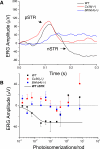
A: in zone I (<0.1 photoisomerizations/rod), very dim stimuli were used to elicit positive and negative scotopic threshold responses (pSTRs and nSTRs). B: all mice had a positive-going response in zone 1; however, only WT mice had nSTR, which can also be fit with an exponential equation.
FIG. 5.
FIG. 5.
Analysis of the b-wave reveals 2 components: a sensitive pSTR shown as a dashed line and a less sensitive PII component, shown as solid lines. WT mice had a robust pSTR, which could be fit by an exponential function (see Table 2), whereas responses of Cx36(−/−) and Bhlhb4(−/−) mice in the range that produced the pSTR were not fit to the exponential function. For PII, WT and Cx36(−/−) had very similar maximum amplitudes and sensitivity (see Table 1).
FIG. 6.
FIG. 6.
Schematic diagram of the 6 rod and cone synaptic pathways mediating various ERG b-wave components in dark-adapted mouse retina. R, rod; C, cone; DBCR, rod depolarizing bipolar cell; DBCC1, high-sensitivity cone depolarizing bipolar cell; DBCC2, low-sensitivity cone depolarizing bipolar cell; AII, AII amacrine cell; ONGCs: on ganglion cells; thick red arrows (±: sign-preserving/sign-inverting), glutamatergic synapses; red zigzags, electrical synapses; PRL, photoreceptor layer; OPL, outer plexiform layer; INL, inner nuclear layer; IPL, inner plexiform layer; GCL, ganglion cell layer.

References

    1. Applebury ML, Antoch MP, Baxter LC, Chun LL, Falk JD, Farhangfar F, Kage K, Krzystolik MG, Lyass LA, Robbins JT. The murine cone photoreceptor: a single cone type expresses both S and M opsins with retinal spatial patterning. Neuron 27: 513–523, 2000. - PubMed
    1. Bramblett DE, Pennesi ME, Wu SM, Tsai MJ. The transcription factor Bhlhb4 is required for rod bipolar cell maturation. Neuron 43: 779–793, 2004. - PubMed
    1. Bui BV, Fortune B. Ganglion cell contributions to the rat full-field electroretinogram. J Physiol 555: 153–173, 2004. - PMC - PubMed
    1. Calvert PD, Krasnoperova NV, Lyubarsky AL, Isayama T, Nicolo M, Kosaras B, Wong G, Gannon KS, Margolskee RF, Sidman RL, Pugh EN Jr, Makino CL, Lem J. Phototransduction in transgenic mice after targeted deletion of the rod transducin alpha-subunit. Proc Natl Acad Sci USA 97: 13913–13918, 2000. - PMC - PubMed
    1. Carter-Dawson LD, LaVail MM. Rods and cones in the mouse retina. I. Structural analysis using light and electron microscopy. J Comp Neurol 188: 245–262, 1979. - PubMed

Publication types

MeSH terms

LinkOut - more resources