

Visual field maps, population receptive field sizes, and visual field coverage in the human MT+ complex
- PMID: 19587323
- PMCID: PMC2777836
- DOI: 10.1152/jn.00102.2009
Visual field maps, population receptive field sizes, and visual field coverage in the human MT+ complex
Abstract
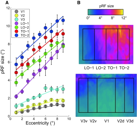
Human neuroimaging experiments typically localize motion-selective cortex (MT+) by contrasting responses to stationary and moving stimuli. It has long been suspected that MT+, located on the lateral surface at the temporal-occipital (TO) boundary, contains several distinct visual field maps, although only one coarse map has been measured. Using a novel functional MRI model-based method we identified two maps-TO-1 and TO-2-and measured population receptive field (pRF) sizes within these maps. The angular representation of the first map, TO-1, has a lower vertical meridian on its posterior side at the boundary with the lateral-occipital cortex (i.e., the LO-2 portion). The angular representation continues through horizontal to the upper vertical meridian at the boundary with the second map, TO-2. The TO-2 angle map reverses from upper to lower visual field at increasingly anterior positions. The TO maps share a parallel eccentricity map in which center-to-periphery is represented in the ventral-to-dorsal direction; both maps have an expanded foveal representation. There is a progressive increase in the pRF size from V1/2/3 to LO-1/2 and TO-1/2, with the largest pRF sizes in TO-2. Further, within each map the pRF size increases as a function of eccentricity. The visual field coverage of both maps extends into the ipsilateral visual field, with larger sensitivity to peripheral ipsilateral stimuli in TO-2 than that in TO-1. The TO maps provide a functional segmentation of human motion-sensitive cortex that enables a more complete characterization of processing in human motion-selective cortex.
Figures


References
-
- Allman JM, Kaas JH. A representation of the visual field in the caudal third of the middle tempral gyrus of the owl monkey (Aotus trivirgatus). Brain Res 31: 85–105, 1971 - PubMed
-
- Ashburner J, Friston K. Rigid body registration. In: Human Brain Function ( 2nd ed.), edited by Frackowiak RSJ, Friston KJ, Frith C, Dolan R, Friston KJ, Price CJ, Zeki S, Ashburner J, Penny WD. San Diego, CA: Academic Press/Elsevier Science, 2003, p. 635–654
-
- Bandettini PA, Jesmanowicz A, Wong EC, Hyde JS. Processing strategies for time-course data sets in functional MRI of the human brain. Magn Reson Med 30: 161–173, 1993 - PubMed
-
- Boussaoud D, Ungerleider LG, Desimone R. Pathways for motion analysis: cortical connections of the medial superior temporal and fundus of the superior temporal visual areas in the macaque. J Comp Neurol 296: 462–495, 1990 - PubMed
Publication types
MeSH terms
Substances
Grants and funding
LinkOut - more resources
Full Text Sources