. 2009 Aug 27;114(9):1974-86.
doi: 10.1182/blood-2009-03-208900. Epub 2009 Jul 8.

Endothelial cell protein C receptor cellular localization and trafficking: potential functional implications

Affiliations

Endothelial cell protein C receptor cellular localization and trafficking: potential functional implications

Ramesh C Nayak et al. Blood. .

Abstract

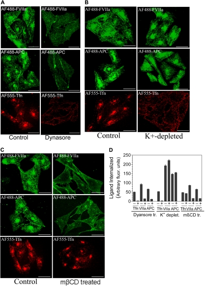
Although the binding of endothelial cell protein C receptor (EPCR) to its ligands is well characterized at the biochemical level, it remains unclear how EPCR interaction with its ligands at the cell surface impacts its cellular trafficking. We characterized the cellular localization and trafficking of EPCR in endothelial cells and a heterologous expression system. Immunofluorescence confocal microscopy studies revealed that a majority of EPCR is localized on the cell surface in membrane microdomains that are positive for caveolin-1. A small fraction of EPCR is also localized intracellularly in the recycling compartment. Factor VIIa (FVIIa) or activated protein C binding to EPCR promoted the internalization of EPCR. EPCR and EPCR-bound ligands were endocytosed rapidly via a dynamin- and caveolar-dependent pathway. The endocytosed receptor-ligand complexes were accumulated in a recycling compartment before being targeted back to the cell surface. EPCR-mediated FVIIa endocytosis/recycling also resulted in transport of FVIIa from the apical to the basal side. In vivo studies in mice showed that blockade of EPCR with EPCR-blocking antibodies impaired the early phase of FVIIa clearance. Overall, our results show that FVIIa or activated protein C binding to EPCR promotes EPCR endocytosis, and EPCR-mediated endocytosis may facilitate the transcytosis of FVIIa and its clearance from the circulation.


Figures

Figure 1
Figure 1
Cellular distribution of EPCR. (A) Confluent monolayers of HUVECs, nonpermeabilized or permeabilized with 0.1% Triton X-100 for 10 minutes, were immunostained with EPCR mAbs (JRK1500, 10 μg/mL), followed by Rhodamine Red-conjugated anti–mouse IgG. Immunofluorescence was analyzed by confocal microscopy. (Left) Images from a single plane of z-stack. (Right) Three-dimensional reconstructed composite images of all z-stacks. (B) CHO-EPCR cells were immunostained with EPCR mAbs as described in panel A. (C) HUVECs (top panel) and CHO-EPCR cells (bottom panel) were first treated with either control vehicle or mβCD (10 mM) for 30 minutes (to deplete the membrane cholesterol) and then permeabilized with 0.1% Triton X-100 for 10 minutes. The permeabilized cells were immunostained with polyclonal anti–human caveolin-1 and EPCR mAbs followed by Oregon Green–labeled anti–rabbit IgG and Rhodamine Red-labeled anti–mouse IgG as secondary reporter antibodies. (Right, merge) Overlay of caveolin-1 and EPCR staining. The images shown were composite images. (D) CHO cells were transfected transiently with full-length EPCR-GFP or EPCR lacking N-terminal signal peptide (EPCR Δsp)–GFP fusion construct, and the expression of green fluorescent fusion product was analyzed by confocal microscopy. (E) CHO-EPCR cells were permeabilized and immunostained with polyclonal anti–γ-tubulin and EPCR mAbs. (Insets) The magnified view of the boxed regions. Arrow represents separation of green and red fluorescence, which indicates that γ-tubulin is not colocalized with EPCR. The presence of EPCR around γ-tubulin indicates that EPCR is localized in the pericentriolar region. (F) CHO-EPCR cells were immunostained either with polyclonal anti-rab11 and EPCR mAb (top panel) or transiently transfected with rab11-GFP and then immunostained with EPCR mAb (bottom). (Insets) Magnified view of the boxed regions. It is known that rab11 also associates with other endosomal compartments and secretory vesicles; therefore, rab11 antibodies, in addition to the REC, also stained other intracellular compartments. (G) CHO-EPCR cells (top panel) or HUVECs (bottom panel) were treated with AF555-transferrin (300 nM) for 1 hour and then immunostained with EPCR mAbs. Arrows in the right panel represent the colocalization of EPCR and AF555-trasferrin in the REC. Scale bar represents 15 μm. The REC is not visual in all cells as its position could vary from cell to cell. The REC that is positioned on top of the nucleus may give a false impression that it is localized in the nucleus.
Figure 2
Figure 2
Intracellular distribution of EPCR in endothelial cells. Permeabilized HUVECs were immunostained with EPCR mAbs and an organelle-specific antibody. (Left panel) EPCR-specific staining. (Middle panel) Organelle-specific staining. (Right panel) The overlay image (colocalization) of organelle-specific marker and EPCR. Organelle-specific antibodies used were as follows: anti-EEA1 and anti-rab5 for early endosomes, anti-LAMP1 for lysosomes, Giantin for the Golgi and rab11 for the recycling compartment. A small portion of the merged image, bordered with the white box, was digitally enlarged to illustrate the colocalization.
Figure 3
Figure 3
Internalization of FVIIa bound to EPCR. (A) HUVECs were exposed to AF488-FVIIa (50 nM) for 15 minutes, 30 minutes, or 1 hour at 37°C, and then fixed and processed for EPCR immunostaining. (Left) EPCR staining. (Middle) AF488-FVIIa fluorescence. (Right) The overlay image of left and middle panels. Arrows represent the accumulation of AF488-FVIIa with EPCR in the REC. (B) CHO-EPCR cells were exposed to AF488-FVIIa (50 nM) for 1 hour at 4°C. At the end of 1 hour, the supernatant was removed, and the cells were washed quickly with calcium-containing buffer to remove the unbound ligands and then transferred to 37°C to allow internalization of the surface-bound ligands. Various times at 37°C, the cells were fixed, permeabilized, and immunostained with nonblocking EPCR mAb. (Left) AF488-FVIIa. (Middle) EPCR staining. (Right) The merged image of AF488-FVIIa and EPCR. Because the REC could locate at apical, basal, or lateral position to the nucleus, it may not be visible in all sections of the cell. (C) Ligand-induced EPCR accumulation in the REC. CHO-EPCR cells were first incubated with FVIIa (50 nM) at 4°C for 1 hour and then transferred to 37°C. At various times, the cells were fixed, permeabilized, and stained with EPCR mAbs. The pixel density of the fluorescence of EPCR staining in the REC at different time periods was measured using Image suite software (PerkinElmer Life and Analytical Sciences; n = 15 cells or more).
Figure 4
Figure 4
FVIIa or APC binding to EPCR leads to internalization of both the ligand and the receptor. CHO-EPCR cells were incubated with 125I-labeled FVIIa (□) or APC (Δ; 10 nM) for various time intervals at 37°C, and the amount of 125I-labeled proteins associated with the cell surface (A) and internalized (B) was measured. Data are mean ± SEM (n = 4-5). (C) CHO-EPCR cells were surface-labeled with sulfo-NHS-SS-biotin at 4°C and then incubated at 37°C for various time periods with a control buffer or the buffer containing FVIIa or APC (10 nM). The cells were then treated with the reducing agent to remove biotin label from the cell surface, lysed, and immunoprecipitated with anti-EPCR antibody. The immunoprecipitated samples were subjected to SDS-PAGE followed by immunoblot analysis with antibiotin antibodies to detect the internalized EPCR. Biotinylated EPCR signal was quantified by densitometry. Biotinylated EPCR signal detected in the cells immediately after the biotinylation was taken as 100%. The values shown in the figure represent mean ± SEM (n = 3).
Figure 5
Figure 5
EPCR-mediated internalization of FVIIa and APC is dependent on dynamin and caveolar-mediated endocytosis. (A) CHO-EPCR were treated with a control vehicle (0.25% dimethyl sulfoxide) or Dynasore (80 μM), a specific inhibitor of dynamin GTPase, for 30 minutes. (B) CHO-EPCR cells were treated with control or K+-depleted buffer to inhibit clathrin-dependent endocytosis. (C) CHO-EPCR cells were treated with a control vehicle or mβCD (10 mM) for 30 minutes to disrupt caveolae. After aforementioned specific treatments, the cells were exposed to AF488-FVIIa (50 nM), AF488-APC (50 nM), or AF555-transferrin (Tfn; 300 nM) for 1 hour at 37°C. The fluorescence of internalized ligands was analyzed by confocal microscopy. The extent of internalization was quantified (D) by measuring the fluorescence intensity in the defined area corresponding to the recycling compartment. To identify the recycling compartment for the quantification, the cells were immunostained with EPCR mAbs (mean ± SEM, n = 15-25 cells).
Figure 6
Figure 6
Recycling of internalized FVIIa and EPCR, and the fate of internalized ligands. (A) CHO-EPCR cells were incubated with AF488-FVIIa (50 nM) for 1 hour at 37°C to allow the internalization of FVIIa. Thereafter, the cells were washed with the buffer containing 5 mM ethylenediaminetetraacetic acid to remove the cell surface bound FVIIa, and thereafter the cells were maintained in calcium-containing buffer (buffer B) at 37°C. The fate of internalized FVIIa was monitored by fixing the cells at various times. The fixed cells were permeabilized, stained with EPCR mAbs, and analyzed by confocal microscopy. (B-C) CHO-EPCR cells were exposed to 125I-FVIIa (triangle) or 125I-APC (square), 10 nM, for 2 hours at 37°C, and then the surface associated 125I-labeled ligand was eluted by treating the cells with 0.1 M glycine, pH 2.3, for 3 minutes at the room temperature. The cells were washed with buffer B and allowed to stay at 37°C. At various time intervals, the overlying supernatant medium was removed and precipitated with 10% ice-cold TCA. The radioactivity present in both TCA-precipitable (B) and TCA-soluble fractions (C) was counted. (D) CHO-EPCR cells were incubated with a control buffer or the buffer containing unlabeled FVII, FVIIa, protein C, or APC (100 nM). After 2 hours at 37°C, the unbound ligand was removed, and the monolayers were washed with 0.1 M glycine, pH 2.3, to remove the bound ligand. After washing the monolayers with buffer B, the cells were chilled on ice and incubated with 125I-EPCR mAbs (10 nM) at 4°C for 1 hour. After 1 hour, the unbound radioactivity was removed, the cells were washed, and the total cell lysate was counted for the radioactivity to determine the amount of EPCR mAbs bound to the cells.
Figure 7
Figure 7
Involvement of rab11 in sorting the internalized FVIIa into the recycling compartment. CHO-EPCR cells were transiently transfected with wild-type rab11, constitutively active rab11 (Q70L), or dominant negative rab11 (S25N), and the transfected cells were exposed to AF488-FVIIa for 1 hour at 37°C. The cells were fixed, permeabilized, and immunostained for rab11. The fluorescence of internalized AF488-FVIIa and immunofluorescence of rab11 were analyzed by confocal microscopy.
Figure 8
Figure 8
EPCR-mediated FVIIa transcytosis. (A) CHO-EPCR cells cultured in transwells were treated with control vehicle or EPCR blocking mAb (10 μg/mL) for 30 minutes. Thereafter, 125I-FVIIa (10 nM) and BSA coupled to Evans Blue (0.67 mg/mL) were added to the upper chamber, and the cells were allowed to stay in the CO2 incubator. At various times, a small aliquot was removed from the bottom chamber and counted for the radioactivity and measured absorbance at 650 nm to monitor the transfer of FVIIa and BSA, respectively, to the bottom chamber. The data shown in the figure represent EPCR-specific FVIIa transport (n = 3). BSA transfer in CHO-EPCR cells treated with control vehicle or EPCR blocking mAbs is very similar. (B) C57BL/6 mice were injected with saline control (100 μL), AF488-FVII, or AF488-protein C (10 μg/mice in 100 μL saline) via tail vein. One hour after the injection, the mice were exsanguinated, and various tissues were collected into Excel fixative. Bone-joint tissue was sectioned (5-μm thickness) and immunostained with anti-AF488 antibodies to localize the administered FVII and protein C. [↓] represents endothelial lining; [formula image], adventitia and extravascular tissue.

References

    1. Regan LM, Stearns-Kurosawa DJ, Kurosawa S, et al. The endothelial cell protein C receptor: inhibition of activated protein C anticoagulant function without modulation of reaction with proteinase inhibitors. J Biol Chem. 1996;271(29):17499–17503. - PubMed
    1. Laszik Z, Mitro A, Taylor FB, Jr, Ferrell G, Esmon CT. Human protein C receptor is present primarily on endothelium of large blood vessels: implications for the control of the protein C pathway. Circulation. 1997;96(10):3633–3640. - PubMed
    1. Stearns-Kurosawa DJ, Kurosawa S, Mollica JS, Ferrell GL, Esmon CT. The endothelial cell protein C receptor augments protein C activation by the thrombin-thrombomodulin complex. Proc Natl Acad Sci U S A. 1996;93(19):10212–10216. - PMC - PubMed
    1. Esmon CT. The endothelial protein C receptor. Curr Opin Hematol. 2006;13(5):382–385. - PubMed
    1. Riewald M, Petrovan RJ, Donner A, Mueller BM, Ruf W. Activation of endothelial cell protease activated receptor 1 by protein C pathway. Science. 2002;296(5574):1880–1882. - PubMed

Publication types