. 2009 Aug;119(8):2330-42.
doi: 10.1172/JCI39087. Epub 2009 Jul 1.

CSF-1 signals directly to renal tubular epithelial cells to mediate repair in mice

Affiliations

CSF-1 signals directly to renal tubular epithelial cells to mediate repair in mice

Julia Menke et al. J Clin Invest. 2009 Aug.

Abstract

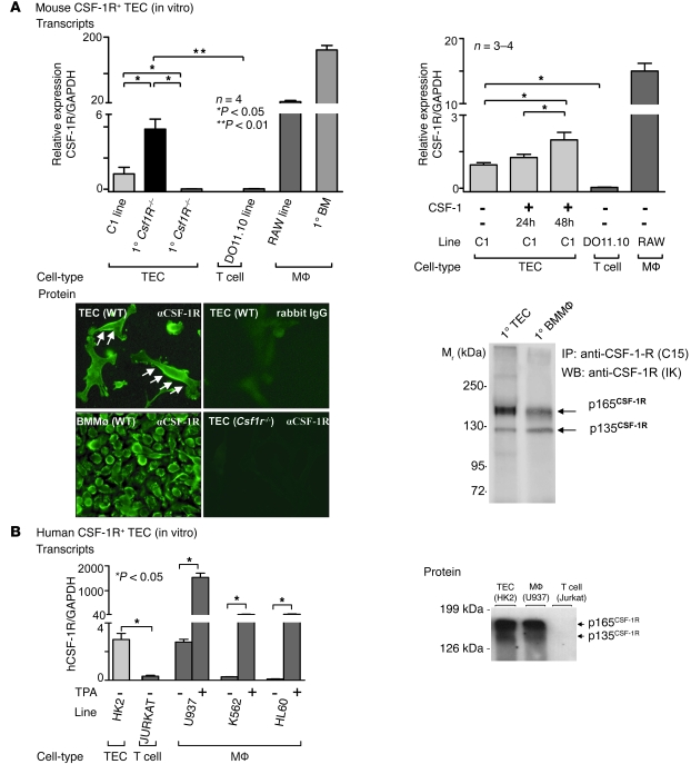
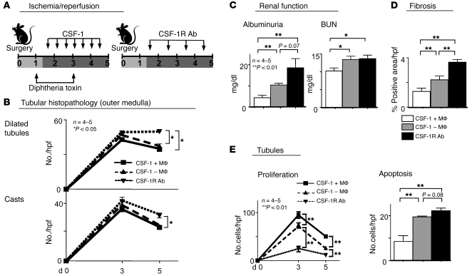
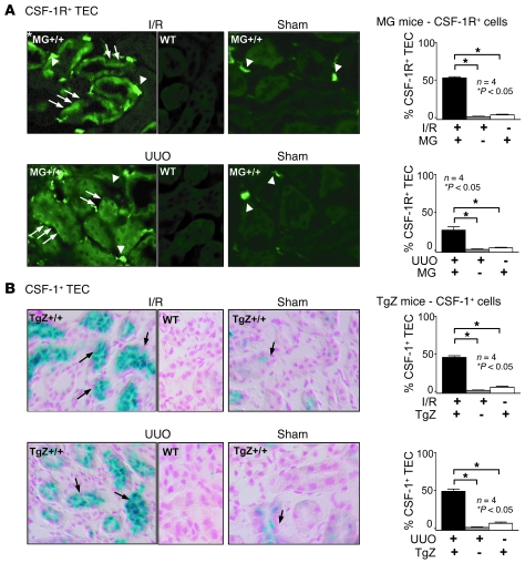
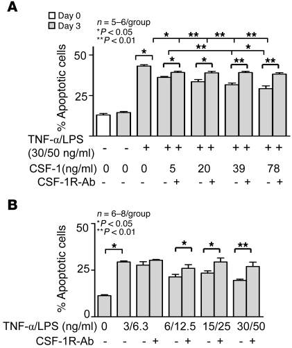
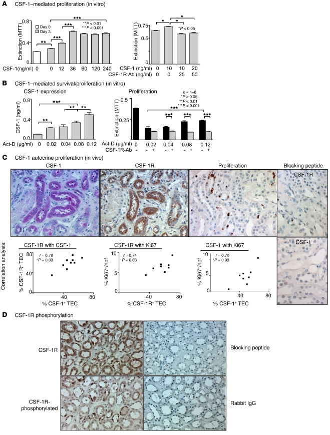
Tubular damage following ischemic renal injury is often reversible, and tubular epithelial cell (TEC) proliferation is a hallmark of tubular repair. Macrophages have been implicated in tissue repair, and CSF-1, the principal macrophage growth factor, is expressed by TECs. We therefore tested the hypothesis that CSF-1 is central to tubular repair using an acute renal injury and repair model, ischemia/reperfusion (I/R). Mice injected with CSF-1 following I/R exhibited hastened healing, as evidenced by decreased tubular pathology, reduced fibrosis, and improved renal function. Notably, CSF-1 treatment increased TEC proliferation and reduced TEC apoptosis. Moreover, administration of a CSF-1 receptor-specific (CSF-1R-specific) antibody after I/R increased tubular pathology and fibrosis, suppressed TEC proliferation, and heightened TEC apoptosis. To determine the contribution of macrophages to CSF-1-dependent renal repair, we assessed the effect of CSF-1 on I/R in mice in which CD11b+ cells were genetically ablated and determined that macrophages only partially accounted for CSF-1-dependent tubular repair. We found that TECs expressed the CSF-1R and that this receptor was upregulated and coexpressed with CSF-1 in TECs following renal injury in mice and humans. Furthermore, signaling via the CSF-1R stimulated proliferation and reduced apoptosis in human and mouse TECs. Taken together, these data suggest that CSF-1 mediates renal repair by both a macrophage-dependent mechanism and direct autocrine/paracrine action on TECs.


Figures

Figure 1
Figure 1. CSF-1 hastens kidney repair and blocking CSF-1R hinders kidney repair following I/R.
(A) Left panel: B6 mice were injected twice daily with either CSF-1 or PBS starting at day 1.5 following I/R until day 4.5. Right panel: B6 mice were injected daily with either CSF-1R or an irrelevant control Ab (rat IgG) starting at day 1.5 following I/R until day 4.5. Except for those shown in part E, all mice were sacrificed for analysis on day 5 following I/R. (B) Left panel: renal tubular histopathology (cortex and outer medulla) was less severe in CSF-1–injected than in PBS-injected mice. Right panel: renal tubular histopathology was more severe in CSF-1R Ab–injected than in control Ab–injected mice. Representative photomicrographs and corresponding graphs indicate the number of dilated tubules and casts in the cortex and outer medulla. (C) Left panel: fibrosis (collagen staining, sirius red) of TECs is decreased in mice receiving CSF-1 compared with those receiving PBS. Right panel: fibrosis is increased in CSF-1R Ab–injected compared with control Ab–injected mice. (D) Left panel: renal function (albuminuria, BUN) is attenuated following I/R in CSF-1–treated compared with PBS-treated B6 mice. Right panel: CSF-1R Ab treatment resulted in a rise of albuminuria. (E) Left panel: mice injected with CSF-1 compared with PBS sacrificed on days 1, 3, and 5 following I/R revealed that TEC proliferation in CSF-1–injected mice peaks and declines more rapidly than in PBS-injected mice. The number of apoptotic TECs evaluated by immunostaining for cleaved caspases-3 decreased in mice receiving CSF-1 compared with those receiving PBS. Right panel: TEC proliferation is reduced while the number of apoptotic cells is increased in CSF-1R Ab–injected compared with control Ab–injected mice. Representative photomicrographs are shown. Data show means ± SEM. Original magnification, ×40.
Figure 2
Figure 2. CSF-1R expression on mouse and human TECs.
(A) Left panel: mouse TECs express CSF-1R mRNA. CSF-1R transcript expression, normalized to GAPDH expression, determined by real-time PCR in primary cultured TECs derived from WT B6 mice and in cells of the proximal TEC line, C1. TECs from Csf1R–/– mice and a T cell line (DO11.10) served as negative controls, and WT BM macrophages and cells of the RAW 264 macrophage cell line served as positive controls. Right panel: CSF-1 upregulates TEC CSF-1R transcript expression. C1 cells were incubated with CSF-1 (100 ng/ml) for 24 hours and 48 hours. Results are representative of 3 separate experiments. Means ± SEM are shown. Bottom left panel: Primary TECs express CSF-1R protein. Primary WT BM macrophages and primary Csf1R–/– TECs served as positive and negative controls, respectively (n = 2–3 per group). Original magnification, ×40. Bottom right panel: CSF-1R protein detected in lysates of cultured primary WT TECs. Western blot of CSF-1R immunoprecipitates. WT BM macrophages (at less than one-third the amount of TEC protein) served as a positive control. (B) Left panel: CSF-1R transcript expression in cultured cells of the human proximal TEC line, HK2, determined by real-time PCR. The Jurkat T cell line, the human leukemic monocytic leukemia (U937), the human erythromyeloblastoid leukemia cell line (K562), and the human leukemic cell line (HL60), all stimulated with TPA, served as positive controls. Results are representative of 3 separate experiments. Data represent means ± SEM. Right panel: Human TECs (HK2 line) express CSF-1R protein detected by Western blotting. TPA-stimulated U937 cells and Jurkat T cells served as positive and negative controls, respectively.
Figure 3
Figure 3. Macrophages are only partially responsible for CSF-1–dependent renal repair.
(A) CD11b-DTR and WT mice were injected with CSF-1 following renal injury. We injected DT to deplete macrophages as indicated in the diagram, and we injected anti–CSF-1R Abs as in the diagram. (B) Tubular pathology (outer medulla) and (E) TEC proliferation were analyzed at days 0, 3, and 5 after I/R. (C) Renal function (albuminuria, BUN), (D) fibrosis (collagen), and (E) tubular apoptosis (caspase-3) were analyzed at day 5 after I/R. Data represent means ± SEM.
Figure 4
Figure 4. CSF-1R and CSF-1 expression on TECs are increased following renal injury.
(A) CSF-1R gene expression is upregulated on TECs following I/R and UUO. TECs derived from MacGreen;B6 (MG+/+;B6) mice have increased EGFP (arrows) 3 days after I/R or 5 days after unilateral ureter ligation (UUO) as compared with those derived from sham control and WT kidneys. Note EGFP+ macrophages in the renal interstitium (arrowheads). Original magnification, ×40. Right panels: representative photomicrographs from MacGreen;B6 kidneys after I/R or UUO, together with quantitation of the percentage of EGFP+ TECs. (B) CSF-1 gene expression is upregulated on TECs following I/R and UUO. CSF-1 reporter lacZ expression by TECs in TgZ;B6 mice detected by X-gal staining 3 days after I/R or 5 days after UUO as compared with that in sham controls and WT kidneys. Representative photomicrographs. Original magnification, ×40. Right panels: quantitation of the percentage of β-gal–positive TECs. Data show means ± SEM.
Figure 6
Figure 6. CSF-1 protects TECs by dampening apoptosis following injury.
(A) CSF-1 suppresses TEC apoptosis induced by a 72-hour treatment of TECs with TNF-α/LPS. CSF-1 reduces TNF-α/LPS–induced TEC apoptosis assessed by flow cytometric annexin-PI analysis. Blocking the CSF-1R prevents CSF-1–mediated reduction in the number of apoptotic TECs. (B) TNF-α /LPS–injured TECs are self protective through a CSF-1–dependent mechanism that dampens TEC apoptosis. H2K cells were cultured for 72 hours in the absence of CSF-1. Blocking the CSF-1R increases TNF-α/LPS–induced TEC apoptosis. Data show means ± SEM. n = 5–6 per group.
Figure 5
Figure 5. CSF-1 autocrine/paracrine regulated proliferation of human TECs.
(A) Left panel: CSF-1 mediates HK2 TEC proliferation. CSF-1 stimulates proliferation of human HK2 TECs. Right panel: anti–CSF-1R Abs suppress CSF-1–mediated HK2 TEC proliferation. MTT assay. (B) Left panel: TEC injury increases TEC secretion of CSF-1. Following exposure of cultured HK2 TECs to increasing concentrations of actinomycin D (act-D) for 72 hours, we analyzed CSF-1 in the supernatant by ELISA. Right panel: anti–CSF-1R Abs inhibit TEC survival/proliferation in response to TEC injury. HK2 TECs were incubated with the indicated concentrations of actinomycin D in the presence and absence of anti–CSF-1R Abs for 72 hours prior to determination of cell mass by MTT assay. (C) CSF-1 expression, CSF-1R expression, and proliferation of TECs correlate in human transplants with tubular injury. Using serial sections of kidney biopsy specimens from patients with tubular injury and impaired renal function, we probed for CSF-1, CSF-1R, and proliferation in tubules. CSF-1R and CSF-1 are coexpressed on TECs by immunostaining. Note proliferating Ki67+ TECs in tubules coexpressing CSF-1 and CSF-1R. Right panels: CSF-1R and CSF-1 Ab specificity was verified using peptide preabsorption. Representative photomicrographs and correlation graphs. Original magnification, ×20. Data show means ± SEM. n = 8 per group. (D) CSF-1R is tyrosine phosphorylated in human transplants with tubular injury. Using serial sections of kidney biopsy specimens, we probed for CSF-1R and CSF-1R phosphorylation at Y723. CSF-1R and tyrosine-phosphorylated CSF-1R are coexpressed on TECs by immunostaining. CSF-1R and phospho-Y723 CSF-1R Ab specificity was verified using peptide preabsorption and rabbit IgG, respectively. Original magnification, ×40.

Similar articles

Cited by

References

    1. Humphreys B.D., et al. Intrinsic epithelial cells repair the kidney after injury. Cell Stem Cell. 2008;2:284–291. doi: 10.1016/j.stem.2008.01.014. - DOI - PubMed
    1. Goligorsky M.S. Immune system in renal injury and repair: burning the candle from both ends? Pharmacol. Res. 2008;58:122–128. doi: 10.1016/j.phrs.2008.05.011. - DOI - PMC - PubMed
    1. Witzgall R., Brown D., Schwarz C., Bonventre J.V. Localization of proliferating cell nuclear antigen, vimentin, c-Fos, and clusterin in the postischemic kidney. Evidence for a heterogenous genetic response among nephron segments, and a large pool of mitotically active and dedifferentiated cells. J. Clin. Invest. 1994;93:2175–2188. doi: 10.1172/JCI117214. - DOI - PMC - PubMed
    1. Thadhani R., Pascual M., Bonventre J.V. Acute renal failure. N. Engl. J. Med. 1996;334:1448–1460. doi: 10.1056/NEJM199605303342207. - DOI - PubMed
    1. Bonventre J.V., Weinberg J.M. Recent advances in the pathophysiology of ischemic acute renal failure. J. Am. Soc. Nephrol. 2003;14:2199–2210. doi: 10.1097/01.ASN.0000079785.13922.F6. - DOI - PubMed

Publication types

Substances