. 2009 Aug;119(8):2388-98.
doi: 10.1172/JCI37607. Epub 2009 Jul 1.

The TLR9-MyD88 pathway is critical for adaptive immune responses to adeno-associated virus gene therapy vectors in mice

Affiliations

The TLR9-MyD88 pathway is critical for adaptive immune responses to adeno-associated virus gene therapy vectors in mice

Jiangao Zhu et al. J Clin Invest. 2009 Aug.

Abstract

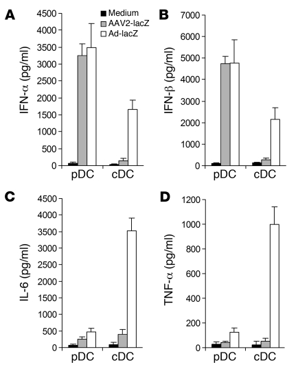
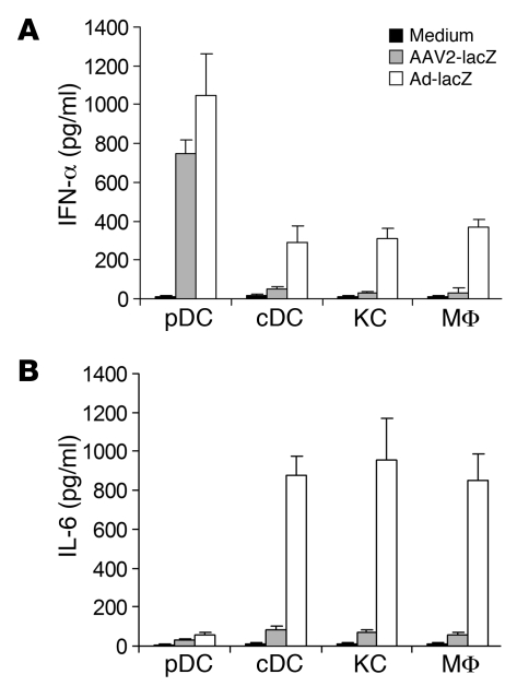
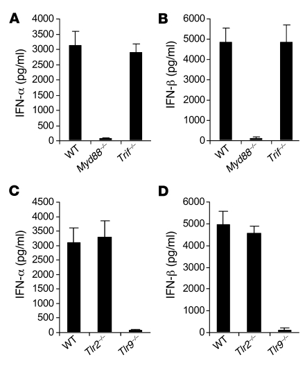
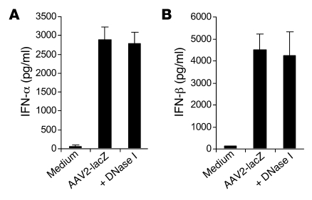
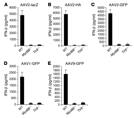
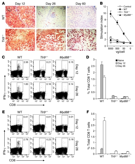
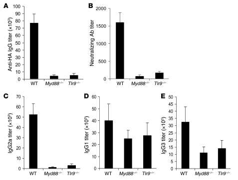
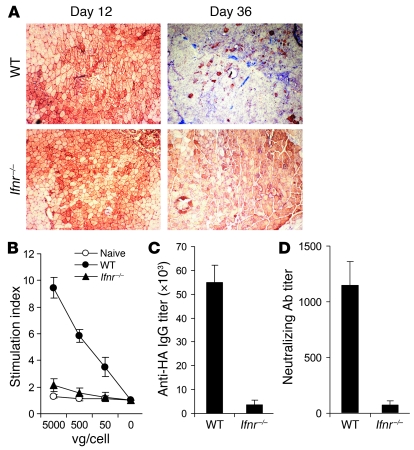
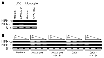
Recombinant adeno-associated viruses (AAVs) have been used widely for in vivo gene therapy. However, adaptive immune responses to AAV have posed a significant hurdle in clinical application of AAV vectors. Recent advances have suggested a crucial role for innate immunity in shaping adaptive immune responses. How AAV activates innate immunity, and thereby promotes AAV-targeted adaptive immune responses, remains unknown. Here we show that AAV activates mouse plasmacytoid DCs (pDCs) via TLR9 to produce type I IFNs. In vivo, the TLR9-MyD88 pathway was crucial to the activation of CD8+ T cell responses to both the transgene product and the AAV capsid, leading to loss of transgene expression and the generation of transgene product-specific and AAV-neutralizing antibodies. We further demonstrate that TLR9-dependent activation of adaptive immunity targeting AAV was mediated by type I IFNs and that human pDCs could be activated in vitro to induce type I IFN production via TLR9. These results reveal an essential role for the TLR9-MyD88-type I IFN pathway in induction of adaptive immune responses to AAV and suggest that strategies that interfere with this pathway may improve the outcome of AAV-mediated gene therapy in humans.


Figures

Figure 1
Figure 1. AAV2 mainly stimulates bone marrow–derived pDCs to secrete type I IFNs.
pDCs and cDCs were generated from bone marrow cells in the presence of Flt-3 ligand and GM-CSF, respectively, and purified by FACS sorting. Cells (1 × 10⁶) were then stimulated with AAV2-lacZ (2 × 10¹⁰ vg) or Ad-lacZ (MOI of 250) or left unstimulated (medium) for 18 hours, and the supernatants were assayed for the secretion of IFN-α (A), IFN-β (B), IL-6 (C), and TNF-α (D) by ELISA. Representative data of 3 independent experiments are shown.
Figure 2
Figure 2. AAV2 activates endogenous pDCs, but not non-pDCs, to produce type I IFNs.
Splenic pDCs, cDCs, hepatic Kupffer cells (KC), or peritoneal macrophages (MF) (2.5 × 10⁵ each) were either unstimulated or stimulated with AAV2-lacZ (5 × 10⁹ vg) or Ad-lacZ (MOI of 250) for 18 hours. The cultured supernatants were assayed for the secretion of IFN-α (A) and IL-6 (B). Representative data of 3 independent experiments are shown.
Figure 3
Figure 3. pDC recognition of AAV2 is mediated by TLR9 and dependent on MyD88.
(A and B) pDCs (1 × 10⁶) generated from bone marrow cells of WT, Myd88–/–, or Trif–/– C57BL/6 mice were purified and stimulated with AAV2-lacZ (2 × 10¹⁰ vg) for 18 hours, and the supernatants were assayed for the secretion of IFN-α (A) and IFN-β (B) by ELISA. (C and D) pDCs generated from bone marrow cells of WT, Tlr2–/–, or Tlr9–/– C57BL/6 mice were stimulated with AAV2-lacZ for 18 hours, and the supernatants were assayed for the secretion of IFN-α (C) and IFN-β (D) by ELISA. Representative data of 3 independent experiments are shown.
Figure 4
Figure 4. DNase I treatment does not affect the ability of AAV to stimulate pDCs.
pDCs (1 × 10⁶) generated from bone marrow cells were purified and stimulated with AAV2-lacZ (2 × 10¹⁰ vg) or DNase I–treated (30 minutes at 37°C) AAV2-lacZ for 18 hours, and the supernatants were assayed for the secretion of IFN-α (A) and IFN-β (B) by ELISA. Representative data of 2 independent experiments are shown.
Figure 5
Figure 5. Activation of the TLR9-MyD88 pathway by AAV is independent of the nature of the transgene or AAV serotypes.
pDCs (1 × 10⁶) generated from WT, Myd88–/–, or Tlr9–/– mice were stimulated with 2 × 10¹⁰ vg of AAV2-lacZ (A), AAV2-HA (B), AAV2-GFP (C), AAV1-GFP (D), or AAV9-GFP (E) for 18 hours, and the supernatants were assayed for the secretion of IFN-β by ELISA. Representative data of 3 independent experiments are shown.
Figure 6
Figure 6. Lack of TLR9-MyD88 signaling diminishes CD8+ T cell responses to the AAV capsid and the transgene product and prolongs the transgene expression.
AAV2-HA (1 × 10¹¹ vg) was injected intramuscularly into WT, Tlr9–/–, or Myd88–/– BALB/c mice. (A) After 12, 26, and 60 days, the infected muscles were harvested and analyzed for HA expression by immunohistochemistry. Original magnification, ×100. (B) CD5+ T cells purified from splenocytes at day 26 after infection, along with uninfected WT splenocytes (control), were restimulated with AAV2-HA at 0, 50, 500, or 5,000 vg/cell. Proliferation of AAV-specific T cells was analyzed by 3H-thymidine incorporation. Data reflect the mean ± SD of the stimulation index, which was calculated by dividing 3H counts in cpm in the presence of viral stimulation by those in the absence of stimulation, as a function of different virus doses. (CF) At days 12 and 26 after infection, splenocytes were harvested and stimulated with either AAV2 capsid epitope peptide (C and D) or HA epitope peptide (E and F) for 5 hours and assayed for intracellular IFN-γ secretion by CD8+ T cells. (C and E) The FACS plots show percentages of IFN-γ–producing CD8+ T cells among total CD8+ T cells. (D and F) The mean percentages ± SD of IFN-γ–producing CD8+ T cells among total CD8+ T cells are also shown. Representative results of 3 independent experiments are shown.
Figure 7
Figure 7. The formation of anti-transgene and AAV-neutralizing antibodies is also dependent on the TLR9-MyD88 pathway.
WT, Tlr9–/–, or Myd88–/– mice were injected with AAV2-HA intramuscularly. (A and B) Serum samples were harvested at day 36 for the measurement of anti-HA antibody titer by ELISA (A) as well as neutralizing antibody titers to AAV vectors (B). (CE) Sera were also analyzed for vector-specific IgG2a (C), IgG1 (D), and IgG3 (E) by ELISA. Data reflect the mean ± SD of reciprocal endpoint titers. Data shown are representative of 3 independent experiments.
Figure 8
Figure 8. Type I IFNs play a critical role in adaptive immune responses to AAV.
AAV2-HA was injected intramuscularly into WT or Ifnr–/– mice. (A) After 12 and 36 days, the infected muscles were harvested and analyzed for HA expression by immunohistochemistry. Original magnification, ×100. (B) CD5+ T cells purified from splenocytes at day 36 after infection, along with uninfected WT splenocytes (naive), were restimulated with AAV2-HA at 0, 50, 500, or 5,000 vg/cell. Proliferation of AAV-specific T cells was analyzed by 3H-thymidine incorporation. Data reflect the mean ± SD of the stimulation index, which was calculated by dividing 3H counts in cpm in the presence of viral stimulation by those in the absence of stimulation, as a function of different virus doses. (C and D) Serum samples were harvested at day 36 for the measurement of anti-HA (C) and AAV-neutralizing (D) antibody titers. Data shown are representative of 2 independent experiments.
Figure 9
Figure 9. Activation of human pDCs by AAV is also mediated by TLR9.
(A) Human pDCs or monocytes (1 × 10⁵) were purified from PBMCs and stimulated with AAV2-lacZ (2 × 10⁹ vg) or left unstimulated for 18 hours. Cells were then harvested, and total RNA was treated with DNase I and assayed for the expression of hIFN-α and hIFN-β by RT-PCR. (B) Human pDCs (1 × 10⁵) were either unstimulated or stimulated with AAV2-lacZ (2 × 10⁹ vg) or a TLR9 agonist, CpG-A ODN (5 μg/ml). In some experiments, cells were pre-treated with the TLR9 antagonist H154 ODN (10 μM) for 30 minutes, followed by stimulation with AAV2-lacZ or CpG-A. After 18 hours, cellular RNA was analyzed for the induction of hIFN-α and hIFN-β by semi-quantitative RT-PCR using 5-fold serial dilution of the template. Human ribosomal protein S14 was used as an internal loading control. Data shown are representative of 2 independent experiments.

Similar articles

Cited by

References

    1. Wu Z., Asokan A., Samulski R.J. Adeno-associated virus serotypes: vector toolkit for human gene therapy. Mol. Ther. 2006;14:316–327. doi: 10.1016/j.ymthe.2006.05.009. - DOI - PubMed
    1. Snyder R.O., et al. Correction of hemophilia B in canine and murine models using recombinant adeno-associated viral vectors. Nat. Med. 1999;5:64–70. doi: 10.1038/4751. - DOI - PubMed
    1. Herzog R.W., et al. Long-term correction of canine hemophilia B by gene transfer of blood coagulation factor IX mediated by adeno-associated viral vector. Nat. Med. 1999;5:56–63. doi: 10.1038/4743. - DOI - PubMed
    1. Kay M.A., et al. Evidence for gene transfer and expression of factor IX in haemophilia B patients treated with an AAV vector. Nat. Genet. 2000;24:257–261. doi: 10.1038/73464. - DOI - PubMed
    1. Manno C.S., et al. Successful transduction of liver in hemophilia by AAV-Factor IX and limitations imposed by the host immune response. Nat. Med. 2006;12:342–347. doi: 10.1038/nm1358. - DOI - PubMed

Publication types