Review
. 2009 Jul;119(7):1784-93.
doi: 10.1172/JCI38177. Epub 2009 Jul 1.

Epidermolysis bullosa simplex: a paradigm for disorders of tissue fragility

Affiliations
Review

Epidermolysis bullosa simplex: a paradigm for disorders of tissue fragility

Pierre A Coulombe et al. J Clin Invest. 2009 Jul.

Abstract

Epidermolysis bullosa (EB) simplex is a rare genetic condition typified by superficial bullous lesions that result from frictional trauma to the skin. Most cases are due to dominantly acting mutations in either keratin 14 (K14) or K5, the type I and II intermediate filament (IF) proteins tasked with forming a pancytoplasmic network of 10-nm filaments in basal keratinocytes of the epidermis and in other stratified epithelia. Defects in K5/K14 filament network architecture cause basal keratinocytes to become fragile and account for their trauma-induced rupture. Here we review how laboratory investigations centered on keratin biology have deepened our understanding of the etiology and pathophysiology of EB simplex and revealed novel avenues for its therapy.


Figures
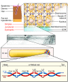
Figure 1
Figure 1. Introduction to the epidermis, keratin filaments, and EB simplex.
Schematic representation of skin tissue and detailed view of the bottom portion of the epidermis, highlighting the cytoplasmic network of keratin IFs attached to hemidesmosome cell-ECM and desmosome cell-cell contacts in basal keratinocytes. The plane of tissue rupture seen in the simplex, junctional, and dystrophic forms of EB are highlighted. The molecular configuration of the K5/K14 coiled-coil heterodimer, the smallest subunit participating in the formation of 10-nm IFs, is shown in a schematic manner. The main secondary structure elements are: N-terminal head and C-terminal tail domains; α-helical coils 1A, 1B, 2A, and 2B; and linkers L1, L12, and L2.
Figure 2
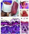
Figure 2. Skin fragility in EB simplex and mouse models thereof.
(A) Example of trauma-induced bullous skin lesions (arrows) in the feet of a 2-month-old child diagnosed with EB simplex. Courtesy of Bernard Cohen (Johns Hopkins School of Medicine; photo copyright DermAtlas). (B) Massive skin blistering as seen in a K14-null mouse neonate. Front paws and facial area are severely affected (arrows). (C) A control littermate exhibits intact skin. (D and E) Micrographs from H&E-stained histological sections prepared from front paws of 2-day-old K14-null (D) and wild-type (E) mice. Fluid-filled blisters are obvious in the K14-null sample. Loss of epidermal integrity occurs near the basal layer of keratinocytes, the defining characteristic of EB simplex. The boxed region in E indicates 3 basal keratinocytes. hf, hair follicles. Scale bar: 100 μm.
Figure 3
Figure 3. Ultrastructure of epidermal basal keratinocytes in EB simplex
These transmission electron micrographs were obtained from ultrathin sections prepared from epoxy-embedded intact (nontraumatized) human skin that had been fixed for routine electron microscopy. (A) Sample from a normal individual. Note the columnar shape of the basal keratinocyte shown and the prominence of keratin IF (KIF) bundles and of dispersed melanin granules (me) in the cytoplasm. (B) Sample from an individual with EBS-DM. Note the two prominent aggregates (Ag) in the cytoplasm, between the nucleus (Nu) and basal lamina (bl). This cell also features KIF bundles, although they seem small compared with those in basal keratinocytes from normal individuals. (C) Sample from an individual with EBS-localized. Note the presence of many vacuoles (vac) in the cytoplasm, between the nucleus and basal lamina in the basal keratinocyte shown. This cell, which does not show obvious defects in KIF content or distribution, may correspond to a microblister. cf, collagen fibers; mi, mitochondria. Scale bar: 1 μm. Adapted from ref. .
Figure 4
Figure 4. Distribution of mutations in K5 and K14 as a function of disease variant in EB simplex.
(A) Histogram depicting the number of cases of mutations in K5 and K14 as a function of EB simplex variant. The category “Others” corresponds to the aggregate of most other variants of the disease, excluding EBS-MD (see Table 1). (B) Distribution of mutations as a function of position within K5 and K14 and clinical variant of the disease. See main text and Figure 1 for information about keratin protein secondary structure. Each entry consists of three numbers: the top one is the total number of mutations; the bottom left number is the subset of these mutations that affect K5; and the bottom right number is the subset of these mutations that affect K14. “None” indicates the absence of mutations for this region of K5 or K14. Red numbers convey the occurrence of a bias toward either K5 or K14. The data are derived from ref. . EBS-AR, EB simplex autosomal recessive; EBS-gen, EBS-generalized; EBS-local, EBS-localized; HIM, helix initiation motif; HTM, helix termination motif.

References

    1. Garg, A., Levin, N.A., and Bernhard, J.D. 2008. Approach to dermatological diagnosis. InFitzpatrick’s dermatology in general medicine. 7th edition. K. Wolf, et al., editors. McGraw-Hill. New York, New York, USA. 23–39.
    1. Fine J.D., et al. Revised classification system for inherited epidermolysis bullosa: report of the Second International Consensus Meeting on diagnosis and classification of epidermolysis bullosa. . J. Am. Acad. Dermatol. 2000;42:1051–1066. - PubMed
    1. Marinkovich, M.P., and Bauer, E.A. 2008. Inherited epidermolysis bullosa. InFitzpatrick’s dermatology in general medicine. 7th edition. K. Wolf, et al., editors. McGraw-Hill. New York, New York, USA. 505–516.
    1. Cooper, T.W., Bauer, E.A., and Briggaman, R.A. 1987. The mechanobullous diseases (epidermolysis bullosa). InFitzpatrick’s dermatology in general medicine. 3rd edition. T.B. Fitzpatrick, et al., editors. McGraw-Hill. New York, New York, USA. 610–626.
    1. Coulombe P.A., Fuchs E. Epidermolysis bullosa simplex. Semin. Dermatol. 1993;12:173–190. - PubMed

Publication types