

Review
doi: 10.1172/JCI38003.
Epub 2009 Jul 1.
Dysfunctions of neuronal and glial intermediate filaments in disease
Affiliations
- PMID: 19587456
- PMCID: PMC2701870
- DOI: 10.1172/JCI38003
Item in Clipboard
Review
Dysfunctions of neuronal and glial intermediate filaments in disease
J Clin Invest.
2009 Jul.
Abstract
Intermediate filaments (IFs) are abundant structures found in most eukaryotic cells, including those in the nervous system. In the CNS, the primary components of neuronal IFs are alpha-internexin and the neurofilament triplet proteins. In the peripheral nervous system, a fifth neuronal IF protein known as peripherin is also present. IFs in astrocytes are primarily composed of glial fibrillary acidic protein (GFAP), although vimentin is also expressed in immature astrocytes and some mature astrocytes. In this Review, we focus on the IFs of glial cells (primarily GFAP) and neurons as well as their relationship to different neurodegenerative diseases.
Figures
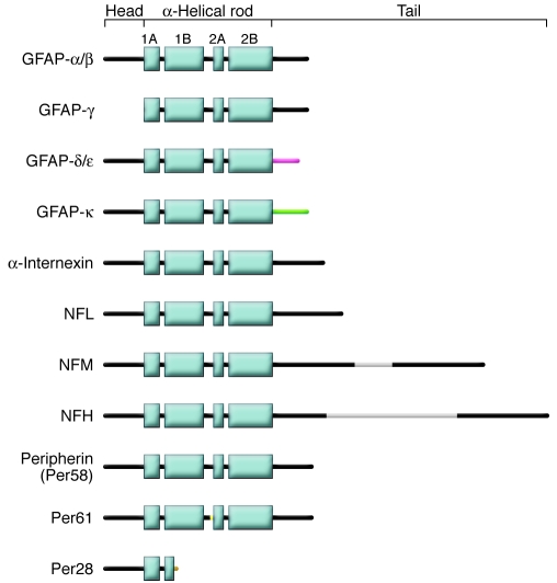
A “typical” IF protein consists of an N-terminal head region, a rod region that contains four α-helical regions (helices 1A, 1B, 2A, and 2B), and a C-terminal tail region. Different isoforms of GFAP are shown: GFAP-α and GFAP-β are full-length GFAP proteins; the mRNAs encoding these two proteins differ in the 5′ UTR. GFAP-γ is encoded by an mRNA that has an alternate start site and is missing exon 1. The mRNAs encoding GFAP-δ, GFAP-ε, and GFAP-κ are generated by alternative splicing of intron 7, with variable use of exon 7+, to create proteins with different C-terminal tail sequences. Only single isoforms of α-internexin, NFL, NFM, and NFH are shown. The gray areas in NFM and NFH contain multiple phosphorylation sites. Full-length peripherin (Per58) is shown as well as Per61, a mouse isoform that retains intron 4 (96), and Per28, which is a mouse and human isoform that retains introns 3 and 4, resulting in a truncated peripherin (97).
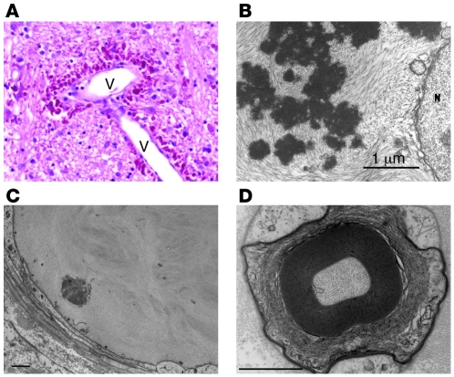
(A) Rosenthal fibers concentrated in the astrocytic endfeet surrounding a blood vessel (V) in the brain stem of a 1-year-old child with Alexander disease. H&E stain, paraffin section (reproduced with permission from Elsevier [102]). Original magnification, ×62. (B) Rosenthal fibers surrounded by IFs in an astrocyte cell body from a 17-month-old child with Alexander disease, viewed by transmission electron microscopy (reproduced with permission of Wiley-Liss Inc., a subsidiary of John Wiley & Sons Inc. [103]). N, nucleus. (C) Sural nerve biopsy from a CMT patient with an L286P mutations in NFL. The figure shows a giant axon with a cluster of organelles (arrow) and irregular whorls of neurofilaments. (D) Sural nerve biopsy of a CMT patient with an NFL del322C–326N mutation. The figure shows a fiber whose axoplasm consists almost exclusively of microtubules; note the loosening of the external myelin lamellae (panels C and D were reproduced with permission from Brain: a journal of neurology [79]). Scale bars: 1 μm (B); 2 μm (C and D).
References
-
- Ching, G.Y., and Liem, R.K.H. 2006. Neuronal intermediate filaments and neurodegenerative diseases. Landes Bioscience. Austin, Texas, USA. 35–51.
-
- Brenner, M., Goldman, J.E., Quinlan, R.A., and Messing, A. 2008. Alexander disease: a genetic disorder of astrocytes. In Astrocytes in pathophysiology of the nervous system. V.H. Parpura and P.G. Haydon, editors. Springer. Boston, Massachusetts, USA. 591–648.
Publication types
MeSH terms
Substances
Grants and funding
LinkOut - more resources
Full Text Sources
Medical
Molecular Biology Databases
Miscellaneous
