Review
. 2009 Jul;119(7):1837-48.
doi: 10.1172/JCI38277. Epub 2009 Jul 1.

Functions of the intermediate filament cytoskeleton in the eye lens

Affiliations
Review

Functions of the intermediate filament cytoskeleton in the eye lens

Shuhua Song et al. J Clin Invest. 2009 Jul.

Abstract

Intermediate filaments (IFs) are a key component of the cytoskeleton in virtually all vertebrate cells, including those of the lens of the eye. IFs help integrate individual cells into their respective tissues. This Review focuses on the lens-specific IF proteins beaded filament structural proteins 1 and 2 (BFSP1 and BFSP2) and their role in lens physiology and disease. Evidence generated in studies in both mice and humans suggests a critical role for these proteins and their filamentous polymers in establishing the optical properties of the eye lens and in maintaining its transparency. For instance, mutations in both BFSP1 and BFSP2 cause cataract in humans. We also explore the potential role of BFSP1 and BFSP2 in aging processes in the lens.


Figures

Figure 1
Figure 1. Schematic of the lens and its cellular organization.
The lens capsule defines the lens perimeter. The anterior of the lens comprises a single layer of epithelial cells, which contains putative stem cells (108). At the lens equator, epithelial cells differentiate into fiber cells (i–iii). Those fiber cells found in the very center of the lens, a region called the lens nucleus, are derived from the posterior epithelial cells of the lens vesicle. Thereafter, the lens grows by the addition of secondary lens fiber cells formed by the differentiation of epithelial cells located at the lens equator. In humans, this begins between weeks 7 and 8 (Carnegie stage 22) after fertilization. This process goes on throughout life in all vertebrates. The epithelial cells differentiate into hexagonal lens fiber cells at the lens equator to maximize cell-cell contacts with neighboring cells (10). It also requires dramatic cell elongation. When corresponding apical and basal ends from opposing fiber cells of the same age from a neighboring lens sector make contact, lens sutures are formed. The geometry of the lens and the arrangement of the lens fiber cells in the human lens into three segments results in Y-shaped sutures, but offset by 60° at each pole. One fiber cell profile has been highlighted (dark blue). Lens fiber cells degrade all their organelles, including mitochondria and nuclei (dots) during differentiation (ii). The bulk of the lens thus consists of long, ribbon-like fiber cells devoid of cytoplasmic organelles (iii). In cross-section, cortical fiber cells show a characteristic hexagonal profile. Diagram adapted with permission from Investigative Ophthalmology & Visual Science (109).
Figure 2
Figure 2. Scanning electron micrographs of bovine lens fiber cells.
The images illustrate the ordered packing of these cells (AC), their hexagonal profile with two longer and four short faces (C) to each cell, and the membrane protrusions at the apex of each short face (B, arrows). Each fiber cell in the outermost layers of the lens is precisely aligned with its neighbors (A, arrows). In cross section, the honeycomb appearance of each fiber cell facilitates tight packing to adjacent cells, thus minimizing intercellular spaces and reducing light scatter. Lens fiber cells are highly elongated, each fiber cell extending from the anterior to posterior pole of the lens, with the length being in excess of 104 μm (see Figure 3). This means that in addition to being elongated, lens fiber cells are also polarized, with distinct apical (anterior-located) and basal (posterior-located) ends. To ensure the correct interdigitation of neighboring fiber cells, there are plasma membrane protrusions concentrated at the apices of the short faces of lens fiber cells (B, arrows). These are most easily seen when the fracture plane has exposed successive layers of fiber cells (B). At high magnification, a regular pattern is seen on the plasma membranes comprising the long faces of the lens fiber cell hexagonal profile. At a molecular level, these surfaces contain the two different plasma membrane complexes, with N-cadherin and cadherin-11 complexes located on the shorter faces and the periplakin, periaxin, ezrin, and desmoyokin complex concentrated along the long face (C) (68). Scale bars: 50 μm, 20 μm, and 20 μm in A, B, and C, respectively.
Figure 3
Figure 3. Assembly and sequence characteristics of BFSP1/2 filaments and native beaded filaments.
(A) The micrograph shows that in vitro, human BFSP2 and bovine BFSP1 form 10-nm filaments when mixed together at a molar ratio of 2:1. The human BFSP2 was produced recombinantly, while the BFSP1 was purified from bovine lenses. In the absence of α-crystallins, the BFSP1 and BFSP2 proteins form 10-nm filaments, supporting their inclusion in the IF superfamily. Both proteins were prepared as described in ref. . (B) This micrograph shows the native filaments present in the lens. Two types are apparent: Smooth, 10-nm filaments (arrowheads) are decorated with α-crystallin particles (small arrows). Beaded filaments (large arrows), which also contain α-crystallin particles, are much more abundant and appear untidy in their association with the supporting filament, to the extent that the filament backbone is almost totally obscured. By comparison, the 10-nm filaments are instantly recognizable despite being decorated with α-crystallin particles. These beaded filaments are the structures first described by Maisel and Perry as being lens specific, and it is from these that filensin and CP49 derive the names BFSP1 and BFSP2, respectively. Scale bars: 500 nm.
Figure 4
Figure 4. Schematic highlighting the unusual features of the protein domains in BFSP1 and BFSP2.
Predicted secondary structure for a typical IF protein (vimentin) compared with that predicted for BFSP2 and BFSP1. IF proteins are composed of an α-helical coiled-coil domain flanked by non-helical N and C termini. The boxes represent α-helical domains, which are separated by non-helical linkers L1, L12, and L2. The yellow boxes at the ends of helix 1A and 2B represent the highly conserved LNDR and TYRKLLEGE motifs, in which mutations usually seriously disturb IF function. These motifs are changed in mammalian BFSP1 (LGER and RYHRIIEIE, respectively) and BFSP2 (LGGC and SYHALLDRE, respectively). The R-to-C change in the LNDR motif of BFSP2 would actually cause disease if found in, for example, glial fibrillary acidic protein (GFAP) (110), and for this reason the motif is colored red in the schematic. In fact, an R113C mutation causes cataract in mice when introduced into vimentin (87). Clearly, it is the context of such a change that will determine whether this alone will abrogate filament function. The rod domain of BFSP1 is predicted to be 2–3 nm shorter than that of other mammalian IF proteins due to its shorter helix 2. BFSP2 lacks a C-terminal tail as also observed for keratin 19, a type 1 IF protein.
Figure 5
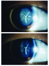
Figure 5. An example of the Y-shaped cataract caused by the E233del BFSP2 mutation.
The cataract is typical of the pedigree recently reported with an E233del BFSP2 mutation (53). Notice the white deposits visible in the lenses of this affected individual taken from a previously published pedigree. The white deposits form a Y shape, which is related to the suture pattern made by the abutting apical and basal ends of the lens fiber cells. The different lighting levels (brighter, top panel; less bright, lower panel) accentuate the anterior (bottom panel) and posterior (top panel) sutures and their associated light scattering deposits. Reproduced with permission from Molecular Vision (53).
Figure 6
Figure 6. Schematic showing the links between beaded filaments and IFs at different plasma membrane sites in the lens fiber cell.
Beaded filaments interact with the plasma membranes of lens fiber cells and are often tightly associated, resisting even alkali extraction (111). Two different protein complexes have been characterized recently in lens fiber cells (68), termed the cadherin- and EPPD-based (ezrin, periplakin, periaxin, desmoyokin–based) complexes, which associate with the short and long faces of fiber cells (see Figure 2), respectively. Plakoglobin and plectin are potential linkers for the IFs and beaded filaments to the cadherin complex. On the long faces of the fiber cells, a number of potential proteins that would link the IFs and beaded filaments to the plasma membrane have been identified (FERM, ankyrin), but the identity of the transmembrane proteins has yet to be determined. Although CD44 homologs were looked for, none were found (68), but the lens does express band 3 (96), which could link to ankyrin and spectrin. Some details of this lens plasma membrane–cytoskeleton complex might parallel that of the erythrocyte.
Figure 7
Figure 7. Summary of the conclusions (blue) and future research directions (green) for IFs and their function in the eye lens.

Similar articles

Cited by

References

    1. Ishikawa H., Bischoff R., Holtzer H. Mitosis and intermediate-sized filaments in developing skeletal muscle. J. Cell Biol. 1968;38:538–555. doi: 10.1083/jcb.38.3.538. - DOI - PMC - PubMed
    1. Hyder C.L., Pallari H.M., Kochin V., Eriksson J.E. Providing cellular signposts — post-translational modifications of intermediate filaments. FEBS Lett. 2008;582:2140–2148. doi: 10.1016/j.febslet.2008.04.064. - DOI - PubMed
    1. Bloemendal H., et al. Ageing and vision: structure, stability and function of lens crystallins. Prog. Biophys. Mol. Biol. 2004;86:407–485. doi: 10.1016/j.pbiomolbio.2003.11.012. - DOI - PubMed
    1. Chepelinsky A.B. Structural function of MIP/aquaporin 0 in the eye lens; genetic defects lead to congenital inherited cataracts. Handb. Exp. Pharmacol. 2009;190:265–297. doi: 10.1007/978-3-540-79885-9_14. - DOI - PubMed
    1. Gong X., Cheng C., Xia C.H. Connexins in lens development and cataractogenesis. J. Membr. Biol. 2007;218:9–12. doi: 10.1007/s00232-007-9033-0. - DOI - PubMed

Publication types