. 2009 Jul 9;4(7):e6186.
doi: 10.1371/journal.pone.0006186.

Oocyte-specific deletion of Pten in mice reveals a stage-specific function of PTEN/PI3K signaling in oocytes in controlling follicular activation

Affiliations

Oocyte-specific deletion of Pten in mice reveals a stage-specific function of PTEN/PI3K signaling in oocytes in controlling follicular activation

Krishna Jagarlamudi et al. PLoS One. .

Abstract

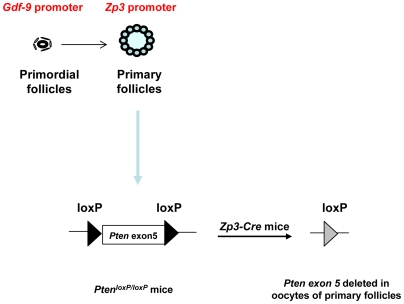
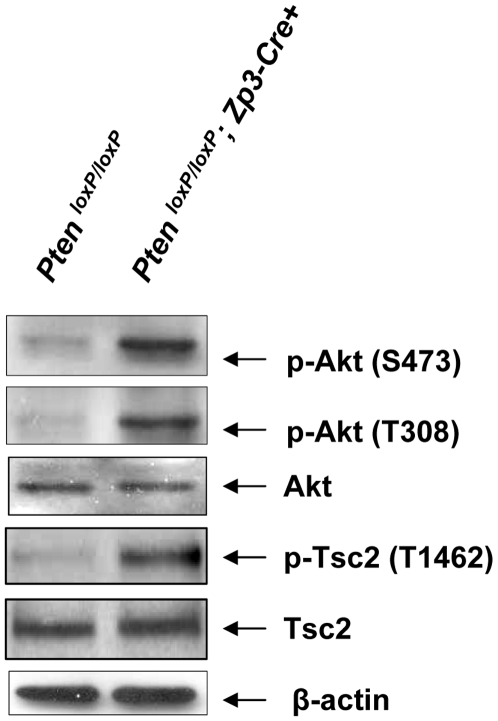
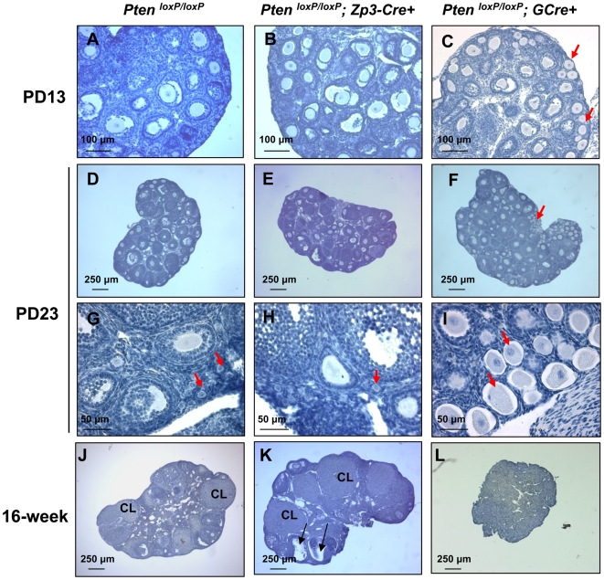
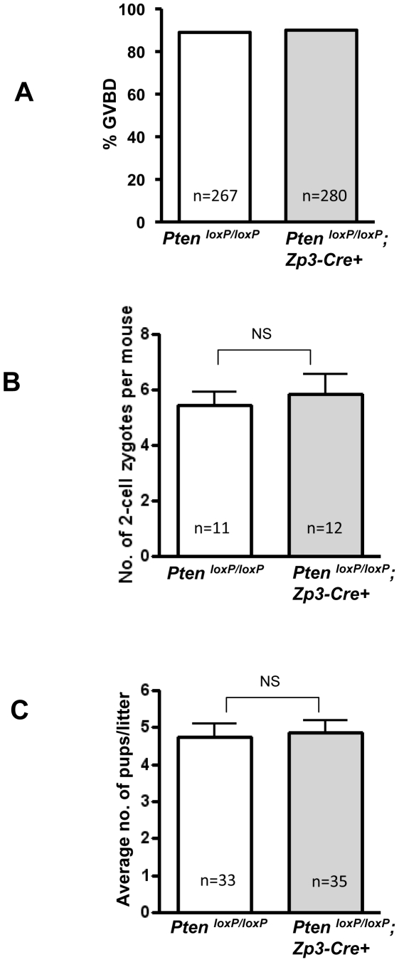
Immature ovarian primordial follicles are essential for maintenance of the reproductive lifespan of female mammals. Recently, it was found that overactivation of the phosphatidylinositol 3-kinase (PI3K) signaling in oocytes of primordial follicles by an oocyte-specific deletion of Pten (phosphatase and tensin homolog deleted on chromosome ten), the gene encoding PI3K negative regulator PTEN, results in premature activation of the entire pool of primordial follicles, indicating that activation of the PI3K pathway in oocytes is important for control of follicular activation. To investigate whether PI3K signaling in oocytes of primary and further developed follicles also plays a role at later stages in follicular development and ovulation, we conditionally deleted the Pten gene from oocytes of primary and further developed follicles by using transgenic mice expressing zona pellucida 3 (Zp3) promoter-mediated Cre recombinase. Our results show that Pten was efficiently deleted from oocytes of primary and further developed follicles, as indicated by the elevated phosphorylation of the major PI3K downstream component Akt. However, follicular development was not altered and oocyte maturation was also normal, which led to normal fertility with unaltered litter size in the mutant mice. Our data indicate that properly controlled PTEN/PI3K-Akt signaling in oocytes is essential for control of the development of primordial follicles whereas overactivation of PI3K signaling in oocytes does not appear to affect the development of growing follicles. This suggests that there is a stage-specific function of PTEN/PI3K signaling in mouse oocytes that controls follicular activation.


Conflict of interest statement

Competing Interests: The authors have declared that no competing interests exist.

Figures

Figure 1
Figure 1. Generation of mutant mice with oocyte-specific deletion of Pten.
A schematic representation of deletion of Pten exon 5 in oocytes of primary and further developed follicles by using the Zp3 promoter-mediated Cre transgenic mice. The developmental stages at which the Gdf-9 promoter and the Zp3 promoter become active are indicated above the illustration of follicles in the figure.
Figure 2
Figure 2. Characterization of Pten deletion by western blot and PCR.
(A) Oocytes were prepared and lysed for western blot as described in Materials and Methods . PTEN expression was found to be completely absent in PtenloxP/loxP; Zp3-Cre+ oocytes. For each lane, 150 oocytes were used. β-actin was used as internal control. (B) PCR analysis showing the complete deletion of Pten exon 5 (Pten Δ5) in one allele of the genomic DNA of pups from PtenloxP/loxP; Zp3-Cre+ females.
Figure 3
Figure 3. Enhanced Akt signaling in oocytes of PtenloxP/loxP; Zp3-Cre+ mice.
Oocytes were prepared from ovaries of 3–4 week old mice that were treated with PMSG, as described in Materials and Methods . Signaling studies in PtenloxP/loxP; Zp3-Cre+ oocytes showed elevated levels of p-Akt (Ser473), p-Akt (Thr308), and p-Tsc2 (Thr1462) as compared to PtenloxP/loxP oocytes. Levels of total Akt, Tsc2, and β-actin were used as internal controls. 100–150 oocytes were used for each lane. All experiments were repeated at least three times and representative results are shown.
Figure 4
Figure 4. Normal follicular development in PtenloxP/loxP; Zp3-Cre+ mice.
Morphological analysis of ovaries from 13- and 23-day-old, and 16-week-old PtenloxP/loxP; Zp3-Cre+ mice, PtenloxP/loxP; GCre+ mice, and control PtenloxP/loxP mice. Ovaries were embedded in paraffin and sections of 8-µm thickness were prepared and stained with hematoxylin. Note the overactivation of primordial follicles in PtenloxP/loxP; GCre+ ovaries (C, F, and I, arrows) and the normal follicular development and CL in PtenloxP/loxP; Zp3-Cre+ ovaries (B, E, H and K), which is comparable to the control PtenloxP/loxP ovaries (A, D, G, and J). CL, corpora lutea.
Figure 5
Figure 5. Normal GVBD, ovulation, fertilization, and fertility in PtenloxP/loxP; Zp3-Cre+ mice.
(A) Oocytes from 3–4-week-old PMSG-primed PtenloxP/loxP; Zp3-Cre+ mice or control PtenloxP/loxP mice were collected in M2 medium. They were cultured further in M16 medium for GVBD analysis. GVBD rate was scored after a culture period of 6 h. Normal GVBD rates were observed in Pten mutant oocytes as compared to control oocytes. The numbers of oocytes used (n) are shown. (B) Two-cell embryos were collected at E1.5 and were cultured in KSOM medium supplemented with amino acids. Numbers of 2-cell embryos per female (mean±SEM) mouse were counted. The numbers of mice used (n) and the results of statistical analysis are shown. No significant difference (NS) was seen between PtenloxP/loxP; Zp3-Cre+ mice and control mice. (C) Normal litter size of PtenloxP/loxP; Zp3-Cre+ females as compared to PtenloxP/loxP control mice (mean±SEM). The numbers of litters (n) from 10 mice of each genotype and results of statistical analysis are shown. NS, not statistically significant.

References

    1. McGee EA, Hsueh AJ. Initial and cyclic recruitment of ovarian follicles. Endocr Rev. 2000;21:200–214. - PubMed
    1. Broekmans FJ, Knauff EA, te Velde ER, Macklon NS, Fauser BC. Female reproductive ageing: current knowledge and future trends. Trends Endocrinol Metab. 2007;18:58–65. - PubMed
    1. Hansen KR, Knowlton NS, Thyer AC, Charleston JS, Soules MR, et al. A new model of reproductive aging: the decline in ovarian non-growing follicle number from birth to menopause. Hum Reprod. 2008;23:699–708. - PubMed
    1. Liu K, Rajareddy S, Liu L, Jagarlamudi K, Boman K, et al. Control of mammalian oocyte growth and early follicular development by the oocyte PI3 kinase pathway: new roles for an old timer. Dev Biol. 2006;299:1–11. - PubMed
    1. Cantley LC. The phosphoinositide 3-kinase pathway. Science. 2002;296:1655–1657. - PubMed

Publication types

Substances