. 2009 Jul 9;4(7):e6180.
doi: 10.1371/journal.pone.0006180.

Improvement of sciatic nerve regeneration using laminin-binding human NGF-beta

Affiliations

Improvement of sciatic nerve regeneration using laminin-binding human NGF-beta

Wenjie Sun et al. PLoS One. .

Abstract

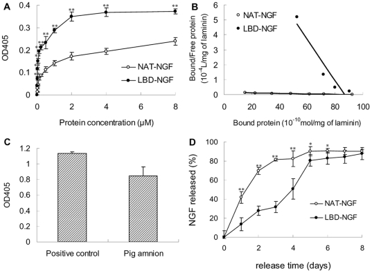
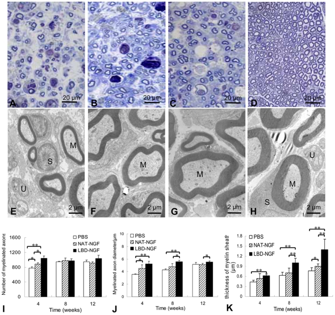
Background: Sciatic nerve injuries often cause partial or total loss of motor, sensory and autonomic functions due to the axon discontinuity, degeneration, and eventual death which finally result in substantial functional loss and decreased quality of life. Nerve growth factor (NGF) plays a critical role in peripheral nerve regeneration. However, the lack of efficient NGF delivery approach limits its clinical applications. We reported here by fusing with the N-terminal domain of agrin (NtA), NGF-beta could target to nerve cells and improve nerve regeneration.

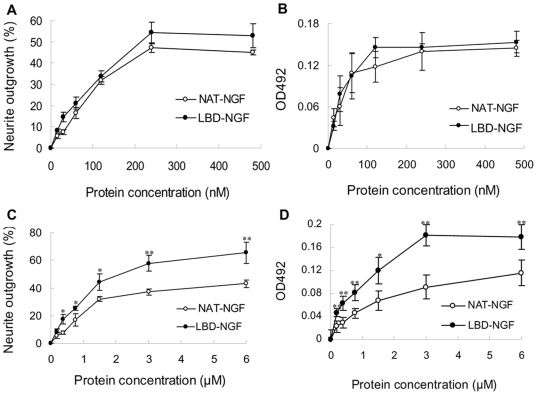
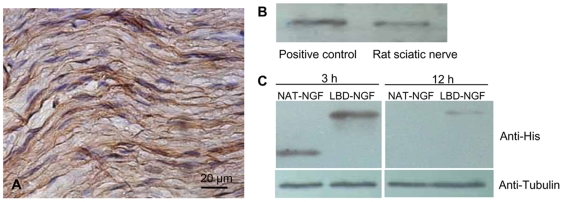
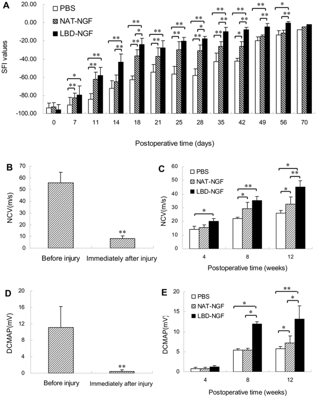
Methods: Laminin-binding assay and sustained release assay of NGF-beta fused with NtA (LBD-NGF) from laminin in vitro were carried out. The bioactivity of LBD-NGF on laminin in vitro was also measured. Using the rat sciatic nerve crush injury model, the nerve repair and functional restoration by utilizing LBD-NGF were tested.

Findings: LBD-NGF could specifically bind to laminin and maintain NGF activity both in vitro and in vivo. In the rat sciatic nerve crush injury model, we found that LBD-NGF could be retained and concentrated at the nerve injury sites to promote nerve repair and enhance functional restoration following nerve damages.

Conclusion: Fused with NtA, NGF-beta could bind to laminin specifically. Since laminin is the major component of nerve extracellular matrix, laminin binding NGF could target to nerve cells and improve the repair of peripheral nerve injuries.


Conflict of interest statement

Competing Interests: The authors have declared that no competing interests exist.

Figures

Figure 1
Figure 1. Laminin-binding and sustained release assay of NAT-NGF and LBD-NGF from laminin in vitro.
(a) Binding curves of NAT-NGF and LBD-NGF to laminin measured by ELISA assay. (b) Kd values for laminin to NAT-NGF and LBD-NGF were calculated using Scatchard analysis. (c) Detection of laminin content in the pig amnion by ELASA assay. (d) Release curves of NAT-NGF and LBD-NGF from laminin in vitro. n = 6, *, P<0.05, **, P<0.01, determined by two-tailed student's t-test.
Figure 2
Figure 2. Bioactivity comparison of NAT-NGF and LBD-NGF in vitro.
(a) Effect of NAT-NGF and LBD-NGF on neurite outgrowth in PC12 cells. (b) Effect of NAT-NGF and LBD-NGF on cell survival in PC12 cells by MTT assay. (c) Percentage of PC12 cells with neurite outgrowth on laminin stimulated by NAT-NGF and LBD-NGF. (d) PC12 cell survival on laminin stimulated by NAT-NGF and LBD-NGF was determined by MTT assay. n = 6, *, P<0.05, **, P<0.01, determined by two-tailed student's t-test.
Figure 3
Figure 3. Detection of laminin content and the sustained NAT-NGF and LBD-NGF in vivo.
(a) Immunohistochemistry of laminin in the rat sciatic nerve. (b) Detection of laminin content in the sciatic nerve by western-blotting analysis. (c) Detection of sustained NAT-NGF and LBD-NGF at the injury sites of sciatic nerves in vivo by western-blotting analysis.
Figure 4
Figure 4. Functional recovery after sciatic nerve injury.
(a) Measurements made from walking track prints were then submitted to SFI. (b) NCV evaluation before and immediately after sciatic nerve injury. (c) NCV evaluation at weeks 4, 8 12 after the sciatic nerve injury. (d) DCMAP evaluation before and immediately after sciatic nerve injury. (e) DCMAP evaluation at weeks 4, 8 12 after the sciatic nerve injury. n = 6, *, P<0.05, **, P<0.01, determined by two-tailed student's t-test.
Figure 5
Figure 5. Histological analysis.
(a–d) At week 8 after injury, the longitudinal sections of the control (a), NAT-NGF (b) and LBD-NGF group (c) compared with that of native nerve group (d) analyzed by H.E. staining. (e–h) At week 8 after injury, immunostaining with anti-neurofilament antibody in transverse sections of the control (e), NAT-NGF (f) and LBD-NGF group (g) compared with that of native nerve group (h). (i–l) At week 8 after injury, immunotaining of the Schwann cell marker S100 in the transverse sections of the control (i), NAT-NGF (j) and LBD-NGF group (k) compared with that of native nerve group (l). (m) The statistical analysis of neurofilament-positive area of each group. (n) The statistical analysis of S100-positive area of each group. n = 6, *, P<0.05, **, P<0.01, determined by two-tailed student's t-test.
Figure 6
Figure 6. Remyelination of sciatic nerves.
(a–d) Toluidine blue staining. At week 4 after injury, light micrographs of transverse semi-thin sections at the injury sites of the control (a), NAT-NGF (b) and LBD-NGF group (c) compared with that of native nerve group (d). (e–h) Transmission electron micrographs (TEMs). At week 12 after injury, ultra-thin sections at the injury sites of the control (a), NAT-NGF (b) and LBD-NGF group (c) compared with that of native nerve group (d) were observed under TEM. (i) The statistical analysis of the number of myelinated axons. (j) The statistical analysis of the myelinated axon diameter. (k) The statistical analysis of thickness of myelin sheath. Myelinated axons (M), unmyelinated axons (U) and Schwann cells (S) surrounding the myelinated axons can be seen clearly. n = 6, *, P<0.05, **, P<0.01, determined by two-tailed student's t-test.

Similar articles

Cited by

References

    1. Kline DG, Kim D, Midha R, Harsh C, Tiel R. Management and results of sciatic nerve injuries: a 24-year experience. J Neurosurg. 1998;89:13–23. - PubMed
    1. Korompilias AV, Payatakes AH, Beris AE, Vekris MD, Afendras GD, et al. Sciatic and peroneal nerve injuries. Microsurgery. 2006;26:288–294. - PubMed
    1. Navarro X, Vivo M, Valero-Cabre A. Neural plasticity after peripheral nerve injury and regeneration. Prog Neurobiol. 2007;82:163–201. - PubMed
    1. Rosberg HE, Carlsson KS, Dahlin LB. Prospective study of patients with injuries to the hand and forearm: costs, function, and general health. Scand J Plast Reconstr Surg Hand Surg. 2005;39:360–369. - PubMed
    1. Lundborg G. A 25-year perspective of peripheral nerve surgery: evolving neuroscientific concepts and clinical significance. J Hand Surg [Am] 2000;25:391–414. - PubMed

Publication types