. 2009 Jul 9;4(7):e6207.
doi: 10.1371/journal.pone.0006207.

Lack of IL-6 during coxsackievirus infection heightens the early immune response resulting in increased severity of chronic autoimmune myocarditis

Affiliations

Lack of IL-6 during coxsackievirus infection heightens the early immune response resulting in increased severity of chronic autoimmune myocarditis

Maya C Poffenberger et al. PLoS One. .

Abstract

Background: Chronic myocarditis is often initiated by viral infection, the most common of which is coxsackievirus infection. The precise mechanism by which viral infection leads to chronic autoimmune pathology is poorly understood, however it is clear that the early immune response plays a critical role. Previous results have shown that the inflammatory cytokine interleukin (IL)-6 is integral to the development of experimental-induced autoimmune myocarditis. However, the function of IL-6 during viral-mediated autoimmunity has yet to be elucidated.

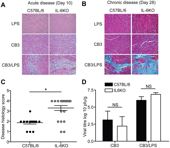
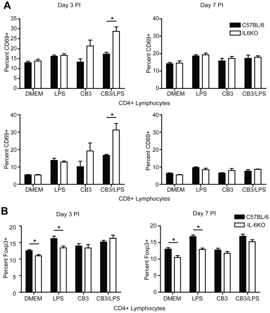
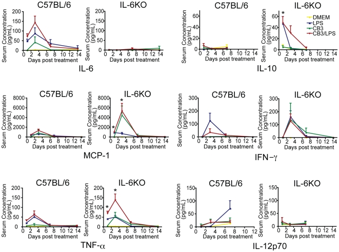
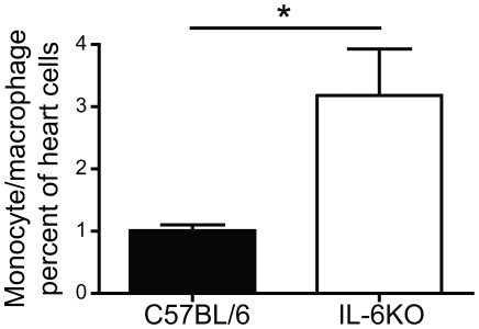
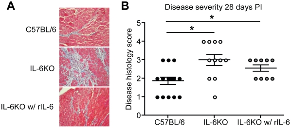
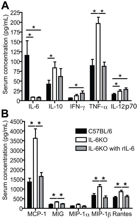
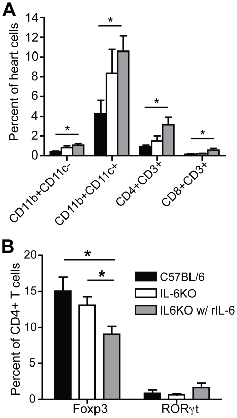
Methods and results: To address the requirement of IL-6 during disease induction, IL-6 deficient mice were infected with coxsackievirus B3 (CB3). Following infection, mice lacking IL-6 developed increased chronic autoimmune disease pathology compared to wild type controls without a corresponding change in the level of viral replication in the heart. This increase in disease severity was accompanied by elevated levels of TNF-alpha, MCP-1, IL-10, activated T cells and cardiac infiltrating macrophage/monocytes. Injection of recombinant IL-6 early following infection in the IL-6 deficient mice was sufficient to lower the serum cytokines TNF-alpha and IL-10 as well as the serum chemokines MCP-1, MIP-1beta, RANTES and MIG with a corresponding decrease in the chronic disease pathology strongly suggests an important regulatory role for IL-6 during the early response.

Conclusions: While IL-6 plays a pathogenic role in experimental-induced autoimmune disease, its function following viral-induced autoimmunity is not reprised. By regulating the early immune response and thereby controlling the severity of chronic disease, IL-6 directs the outcome of chronic autoimmune myocarditis.


Conflict of interest statement

Competing Interests: The authors have declared that no competing interests exist.

Figures

Figure 1
Figure 1. IL-6KO mice develop increased chronic myocarditis severity without an increase in virus in the heart.
(A) Representative Hematoxylin and Eosin stained cardiac sections from C57BL/6 or IL-6KO mice. Mice treated with LPS or CB3 alone did not develop acute disease lesions at day 10 PI (LPS: C57BL/6 n = 4, IL-6KO n = 4) (CB3: C57BL/6 n = 5, IL-6KO n = 4). Mice treated with CB3/LPS developed lesions and immune cell infiltration at 10 days PI (C57BL/6 n = 4, IL-6KO n = 4). Magnification:400×. (B) Representative Masson's Trichrome stained cardiac section from C57BL/6 or IL-6KO mice. Following treatment with CB3/LPS, mice developed disease pathology as determined by fibrosis in blue and immune cell infiltration within the fibrosis areas by 28 days PI (C57BL/6 n = 15, IL-6KO n = 20). Mice infected with LPS or CB3 alone did not develop significant chronic disease pathology (LPS: C57BL/6 n = 5, IL-6KO n = 5) (CB3: C57BL/6 n = 6, IL-6KO n = 7). Magnification:400×. (C) Chronic cardiac disease histology was scored blindly by a four tier grading system to determine severity differences: grade 1, 0–10% pathology; grade 2, 11–25%; grade 3, 26–50%; grade 4, greater than 50% (black circles indicate wt mice, white circles indicate IL-6KO mice) (bar is mean±SE, *p<0.05). Disease severity was found to be significantly higher in the IL-6KO mice compared to wild type controls. (D) Quantification of replicative virus in the heart post infection showed no significant differences in viral titer between IL-6KO and wt mice at day 5 post infection (CB3: C57BL/6 n = 5, IL-6KO n = 5) (CB3/LPS: C57BL/6 n = 5, IL-6KO n = 5) (black bars indicate wt mice, white bars indicate IL-6KO mice) (mean±SE, NS = not significant). This indicates that IL-6 is not essential for control of viral replication in the heart.
Figure 2
Figure 2. IL-6KO mice have increased expression of the early activation marker CD69 at 3 days PI.
(A) T cell activation was monitored by flow cytometry analysis of splenocytes following DMEM, LPS, CB3 or CB3/LPS treatment. Activated T cells were identified by the surface markers CD4, CD8 and CD69. CD69 expression was significantly increased on both CD4+ and CD8+ T lymphocytes at 3 days post infection (DMEM: C57BL/6 n = 4, IL-6KO n = 4) (LPS: C57BL/6 n = 7, IL-6KO n = 8) (CB3: C57BL/6 n = 8, IL-6KO n = 18) (CB3/LPS: C57BL/6 n = 8, IL-6KO n = 17) (black bars indicate wt mice, white bars indicate IL-6KO mice) (mean±SE, *p<0.05) and abrogated by 7 days post infection (DMEM: C57BL/6 n = 4, IL-6KO n = 4) (LPS: C57BL/6 n = 4, IL-6KO n = 4) (CB3: C57BL/6 n = 4, IL-6KO n = 4) (CB3/LPS: C57BL/6 n = 4, IL-6KO n = 5). (B) The percent of CD4+ Treg cells was determined by FACS of splenocytes at 3 and 7 days post DMEM, LPS, CB3 or CB3/LPS treatment. Treg cells were identified by expression of CD4 and the transcription factor Foxp3. IL-6KO mice with DMEM treatment contained a significantly lower percentage of CD4+ Foxp3+ cells compared to the wild type controls with DMEM treatment (DMEM: C57BL/6 n = 8 at day 3 n = 7 at day 7, IL-6KO n = 8 at day 3 n = 7 at day 7) (black bars indicate wt mice, white bars indicate IL-6KO mice) (mean±SE, *p<0.05). However, following treatment with CB3 or CB3/LPS but not LPS alone, the percentage of CD4+ Foxp3+ cells was not significantly different (LPS: C57BL/6 n = 8 at day 3 n = 12 at day 7, IL-6KO n = 8 at day 3 n = 14 at day 7) (CB3: C57BL/6 n = 8 at day 3 n = 11 at day 7, IL-6KO n = 8 at day 3 n = 17 at day 7) (CB3/LPS: C57BL/6 n = 9 at day 3 n = 13 at day 7, IL-6KO n = 9 at day 3 n = 18 at day 7) (mean±SE, *p<0.05) suggesting sufficient proportions of Treg cells are present to immunosuppress disease.
Figure 3
Figure 3. TNF-α, IL-10 and MCP-1 expression is increased in the absence of IL-6.
The serum concentrations of the cytokines IL-12p70, IL-6, IL-10, IFNγ, TNF-α and the chemokine MCP-1 were monitored following treatment with DMEM, LPS, CB3 and CB3/LPS by a BD cytometric bead array inflammation assay (n≥3 for each treatment group at day 1, 3 and 7, n≥2 for each treatment group at day 14) (mean±SE, *p<0.05). IL-6 increased following infection with CB3 indicating a role for IL-6 in the response to viral infection. TNF-α, IL-10 and MCP-1 were significantly increased in the IL-6KO mice compared to the wild type controls indicating a possible role for IL-6 in the regulation of these immune components (orange diamonds indicate DMEM treatment, green squares indicate LPS treatment, blue triangles indicate CB3 treatment, red circles indicate CB3/LPS treatment).
Figure 4
Figure 4. IL-6KO mice have increased monocyte/macrophage infiltration into the heart at 7 days post infection.
Heart infiltrate was measured by flow cytometry analysis at 7 days post treatment with CB3/LPS. Cardiac infiltrate determined based on forward and side scatter as well as CD11b and CD11c immunostaining. IL-6KO mice had significantly increased monocyte/macrophage infiltration at 7 days post treatment compared to wt mice (C57BL/6 n = 9, IL-6KO n = 11) (black bars indicate wt mice, white bars indicate IL-6KO mice) (mean±SE, *p<0.05). This increased correlated with the elevated levels of the chemokine MCP-1 in IL-6KO mice and suggests a link between these two factors.
Figure 5
Figure 5. Injection of rIL-6 in IL-6KO mice decreased chronic disease severity following CB3/LPS treatment.
(A) Representative Masson's Trichrome stained cardiac section from C57BL/6 or IL-6KO mice. Mice were treated with CB3/LPS and either DMEM or rIL-6 on days 0, 1, 2 and 3 post infection. Mice developed disease pathology as determined by fibrosis in blue and immune cell infiltration within the fibrosis areas by 28 days PI (C57BL/6 with DMEM n = 15, IL-6KO with DMEM n = 11, IL-6KO with rIL-6 n = 9). Magnification:400×. (B) Chronic cardiac disease histology was scored blindly by a four tier grading system to determine severity differences: grade 1, 0–10% pathology; grade 2, 11–25%; grade 3, 26–50%; grade 4, greater than 50% (black circles indicate wt mice with DMEM, white circles indicate IL-6KO mice with DMEM, grey circles indicate IL-6KO mice with rIL-6) (bar is mean±SE, *p<0.05). Disease severity was found to be decreased in the IL-6KO mice treated with rIL-6 compared to DMEM treated IL-6KO mice.
Figure 6
Figure 6. Injection of rIL-6 in IL-6KO mice decreased early inflammatory responses at 3 days post infection.
(A) The serum concentrations of the cytokines IL-12p70, IL-6, IL-10, IFNγ and TNF-α were monitored following treatment with CB3/LPS by a BD cytometric bead array inflammation assay at day 3 PI (C57BL/6 with DMEM n = 10, IL-6KO with DMEM n = 9, IL-6KO with rIL-6 n = 10) (black bars indicate wt mice with DMEM, white bars indicate IL-6KO mice with DMEM, grey bars indicate IL-6KO mice with rIL-6) (mean±SE, *p<0.05). TNF-α, IL-10 and IL-12p70 levels were significantly increased in the IL-6KO mice compared to wild type controls. rIL-6 treatment was sufficient to decrease the serum concentration of IL-10 and TNF-α. (B) The serum concentrations of the chemokines MCP-1, MIP-1α, MIP-1β, MIG and RANTES were monitored following treatment with CB3/LPS by a BD cytometric bead array flex set assay at day 3 PI (C57BL/6 n = 10, IL-6KO with DMEM n = 9, IL-6KO with rIL-6 n = 10) (black bars indicate wt mice with DMEM, white bars indicate IL-6KO mice with DMEM, grey bars indicate IL-6KO mice with rIL-6) (mean±SE, *p<0.05). All chemokine levels were increased in the IL-6KO mice compared to wild type controls. Treatment with rIL-6 was sufficient to decrease the levels of all the chemokines.
Figure 7
Figure 7. rIL-6 injection in IL-6KO mice increased innate and adaptive immune cell infiltration in the heart.
(A) Heart infiltrate was measured by flow cytometry analysis at 10 days post treatment with CB3/LPS with or without rIL-6 treatment. Cardiac infiltrate was stained to determine innate and adaptive immune cell infiltration using the surface markers CD11b, CD11c, CD3, CD4 and CD8. rIL-6 treatment in the IL-6KO mice resulted in increased monocyte/macrophages (CD11b+CD11c− and CD11b+CD11c+), CD4+ T cells (CD3+CD4+) and CD8+ T cells (CD3+CD8+) at 10 days post treatment compared to wt and IL-6KO mice treated with DMEM (C57BL/6 with DMEM n = 14, IL-6KO with DMEM n = 10, IL-6KO with rIL-6 n = 11) (black bars indicate wt mice with DMEM, white bars indicate IL-6KO mice with DMEM, grey bars indicate IL-6KO mice with rIL-6) (mean±SE, *p<0.05). (B) The percent of CD4+ cells that are Treg or Th17 cells was determined by flow cytometry analysis of cardiac infiltrate at 10 days post CB3/LPS treatment. Treg cells were identified by the presence of the surface marker CD4 and the transcription factor Foxp3 while Th17 cells were identified by the presence of the surface marker CD4 and the transcription factor RORγt. rIL-6 treated IL-6KO mice contained a significantly lower percentage of CD4+ cells that were Treg cells compared to the DMEM treated wild type controls and DMEM treated IL-6KO mice. Minimal Th17 cells were observed in the heart regardless of treatment (C57BL/6 with DMEM n = 7, IL-6KO with DMEM n = 9, IL-6KO with rIL-6 n = 8) (black bars indicate wt mice with DMEM, white bars indicate IL-6KO mice with DMEM, grey bars indicate IL-6KO mice with rIL-6) (mean±SE, *p<0.05).

Similar articles

Cited by

References

    1. Brown CA, O'Connell JB. Myocarditis and idiopathic dilated cardiomyopathy. Am J Med. 1995;99:309–314. - PMC - PubMed
    1. Eriksson U, Kurrer MO, Schmitz N, Marsch SC, Fontana A, et al. Interleukin-6-deficient mice resist development of autoimmune myocarditis associated with impaired upregulation of complement C3. Circulation. 2003;107:320–325. - PubMed
    1. Kanda T, McManus JE, Nagai R, Imai S, Suzuki T, et al. Modification of viral myocarditis in mice by interleukin-6. Circ Res. 1996;78:848–856. - PubMed
    1. Kanda T, Takahashi T. Interleukin-6 and cardiovascular diseases. Jpn Heart J. 2004;45:183–193. - PubMed
    1. Tanaka T, Kanda T, McManus BM, Kanai H, Akiyama H, et al. Overexpression of interleukin-6 aggravates viral myocarditis: impaired increase in tumor necrosis factor-alpha. J Mol Cell Cardiol. 2001;33:1627–1635. - PubMed

Publication types

MeSH terms