. 2009 Jul 9;4(7):e6203.
doi: 10.1371/journal.pone.0006203.

Cutaneous chronic graft-versus-host disease does not have the abnormal endothelial phenotype or vascular rarefaction characteristic of systemic sclerosis

Affiliations

Cutaneous chronic graft-versus-host disease does not have the abnormal endothelial phenotype or vascular rarefaction characteristic of systemic sclerosis

Jo Nadine Fleming et al. PLoS One. .

Abstract

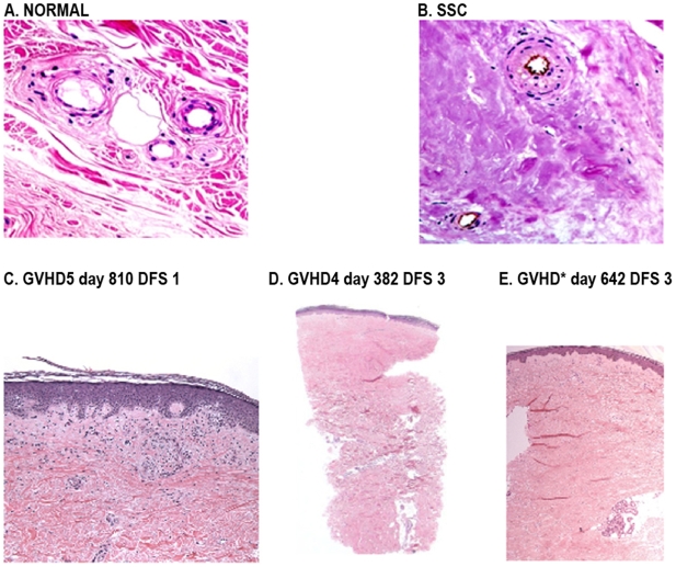
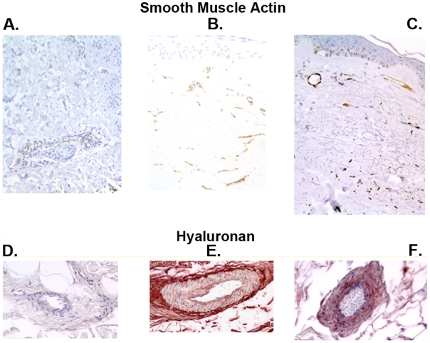
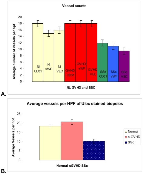
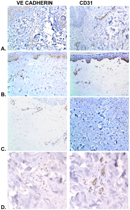
Background: The clinical and histologic appearance of fibrosis in cutaneous lesions in chronic graft-versus -host disease (c-GVHD) resembles the appearance of fibrosis in scleroderma (SSc). Recent studies identified distinctive structural changes in the superficial dermal microvasculature and matrix of SSc skin. We compared the dermal microvasculature in human c-GVHD to SSc to determine if c-GVHD is a suitable model for SSc.

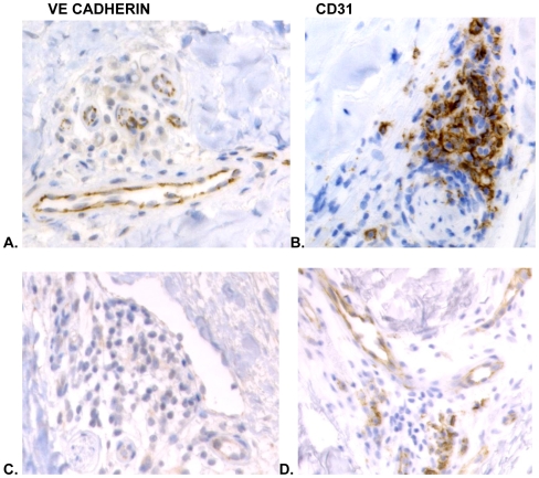
Methodology/principal findings: We analyzed skin biopsies of normal controls (n = 24), patients with SSc (n = 30) and c-GVHD with dermal fibrosis (n = 133)). Immunostaining was employed to identify vessels, vascular smooth muscle, dermal matrix, and cell proliferation. C-GVHD and SSc had similar dermal matrix composition and vascular smooth muscle pathology, including intimal hyperplasia. SSc, however, differed significantly from c-GVHD in three ways. First, there were significantly fewer (p = 0.00001) average vessels in SSc biopsies (9.8) when compared with c-GVHD (16.5). Second, in SSc, endothelial markers were decreased significantly (19/19 and 12/14 for VE cadherin and vWF (p = <0.0001 and <0.05), respectively). In contrast, 0/13 c-GVHD biopsies showed loss of staining with canonical endothelial markers. Third, c-GVHD contained areas of microvascular endothelial proliferation not present in the SSc biopsies.

Conclusions/significance: The sclerosis associated with c-GVHD appears to resemble wound healing. Focal capillary proliferation occurs in early c-GVHD. In contrast, loss of canonical endothelial markers and dermal capillaries is seen in SSc, but not in c-GVHD. The loss of VE cadherin in SSc, in particular, may be related to microvascular rarefaction because VE cadherin is necessary for angiogenesis. C-GVHD is a suitable model for studying dermal fibrosis but may not be applicable for studying the microvascular alterations characteristic of SSc.


Conflict of interest statement

Competing Interests: The authors have declared that no competing interests exist.

Figures

Figure 1
Figure 1. Dermal fibrosis in skin biopsies of Normal, c-GVHD and SSc skin.
Normal skin stained A. with H&E has collagen bundles with curlicue pattern and a dermal fibrosis score (DFS) of zero. 20× magnification B. SSc depicting a DFS of 5 at 20× magnification. C. GVHD day 810 DFS 1: epidermis has patchy lichenoid lymphocytic inflammation along the dermal epidermal junction the widened and papillary dermis contains loosely scattered myofibroblasts and clusters of small proliferated vessels (lower power is 10×) D. GVHD4 day 382 DFS 3: the entire papillary and upper portion of the reticular dermis are replaced by dense fibrosis in a the top-down direction E. GVHD8 day 642 DFS 5: Fibrotic disease throughout the dermis with replacement of the loose papillary dermal collagen and reticular dermal collagen bundles by dense smudgy collagen.
Figure 2
Figure 2. Myofibroblasts and intimal hyperplasia in Normal, GVHD and SSc skin.
Smooth muscle markers were used to depict mural cells and pericytes. A. Smooth muscle actin (SMA) in normal control with normal positive cells in vessels. B. C-GVHD biopsy with many SMA+ myofibroblasts C. Representative SSc biopsy with SMA+ myofibroblasts in dermis slightly lower than c-GVHD Hyaluronan can be used to show abnormally thickened vessels since this marker tends to accumulate on intima and in smooth muscle cells after injury. D. Normal skin showing unaltered vessels in lower dermis. E. C-GVHD biopsy showing thickened vessels with some hyaluronan in the vessel wall. F. Similarly thickened vessels in SSc with increased hyaluronan.
Figure 3
Figure 3. Capillary counts in c-GVHD compared with normal controls and SSc.
Vessel counts by antibody staining A. show SSc has significantly fewer vessels than both other patient groups. Yellow bars show normal tissue vessel counts, red bars show c-GVHD vessels counts, and green blue and purple bars show SSc biopsies stained with CD31, vWF and VE Cadherin respectively. Normal controls and c-GVHD counts are not significantly different from each other regardless of marker used. These counts are also not significantly different within the group (i.e. CD31 stained normal tissue has similar number of vessels to normal tissue stained with vWF). SSc has fewer vessels than c-GVHD and normal tissue with every antibody used to label endothelial cells. In addition, the remaining vessels in SSc (total represented by green bar) a significant proportion of these vessels have lost expression for vWF (blue bar) and VE Cadherin (purple bar). B. Graph shows the results of vascular density quantification after Ulex lectin staining. Normal controls and c-GVHD biopsies are represented by the yellow and orange bars, and are not significantly different. Blue bar represents the average number of vessels in SSc, and is significantly less than the other two groups.
Figure 4
Figure 4. Only SSc has lost endothelial cell markers.
Depicted in A. Normal skin stained with VE cadherin and CD31 showing similar patterns of staining. The same patterns are seen in B. early c-GVHD stained with the same two antibodies and C. late c-GVHD similarly stained with VE cadherin and CD31. The only skin samples which showed loss of VE cadherin were the SSc biopsies D.
Figure 5
Figure 5. Endothelial markers in clumps of microvascular proliferative formations in GVHD but not SSc.
C-GVHD with areas of microvascular proliferation A. Many VE cadherin positive cells present throughout the structure. Some of the endothelial cells within the structure have a normal aggregated stain pattern with VE cadherin at the junctions, wheras in proliferating and migrating endothelial cells the VE cadherin is spread out in the cytoplasm. VE cadherin spread in the cytoplasm is a typical finding in proliferating endothelial cells in glomeruloid bodies in skin. B. CD31 stained biopsies of c-GVHD with glomeruloid bodies with many cells positive for CD31 appearing in large clumps. Similar appearing structures in SSc did not have endothelial markers present in the cells. C. SSc, VE cadherin has few weakly positive cells lining the lumens and very few readily identifiable proliferating endothelial cells D. SSc,CD31 positive cells are sparse in these areas structurally resembling microvascular proliferation. Although multiple lumens are present there are very few CD31 positive clumps of cells.

References

    1. Filipovich AH, Weisdorf D, Pavletic S, Socie G, Wingard JR, Lee S, et al. National Institutes of Health consensus development project on criteria for clinical trials in chronic graft-versus-host disease: I. Diagnosis and staging working group report. Biol Blood Marrow Transplant. 2005;11:945–956. - PubMed
    1. Hymes SR, Turner M, Champlin R, Couriel D. Cutaneous manifestations of chronic graft-versus-host disease. Biol Blood Marrow Transplant. 2006;12:1101–1113. - PubMed
    1. Shulman HM, Kleiner D, Lee SJ, Morton T, Pavletic SZ, Farmer E, et al. Histopathologic diagnosis of chronic graft-versus-host disease: National Institutes of Health Consensus Development Project on Criteria for Clinical Trials in Chronic Graft-versus-Host Disease: II. Pathology Working Group Report. Biol Blood Marrow Transplant. 2006;12:31–47. - PubMed
    1. Shlomchik WD. Graft-versus-host Disease. Nat Rev Immunol. 2007;7:340–352. - PubMed
    1. Zhang Y, McCormick LL, Desai SR, Wu C, Gilliam AC. Murine sclerodermatous graft-versus-host disease, a model for human scleroderma: cutaneous cytokines, chemokines, and immune cell activation. J Immunol. 2002;168:3088–3098. - PubMed

Publication types