Detection and characteristics of microvascular obstruction in reperfused acute myocardial infarction using an optimized protocol for contrast-enhanced cardiovascular magnetic resonance imaging
- PMID: 19588152
- PMCID: PMC2778783
- DOI: 10.1007/s00330-009-1489-0
Detection and characteristics of microvascular obstruction in reperfused acute myocardial infarction using an optimized protocol for contrast-enhanced cardiovascular magnetic resonance imaging
Abstract
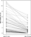
Several cardiovascular magnetic resonance imaging (CMR) techniques are used to detect microvascular obstruction (MVO) after acute myocardial infarction (AMI). To determine the prevalence of MVO and gain more insight into the dynamic changes in appearance of MVO, we studied 84 consecutive patients with a reperfused AMI on average 5 and 104 days after admission, using an optimised single breath-hold 3D inversion recovery gradient echo pulse sequence (IR-GRE) protocol. Early MVO (2 min post-contrast) was detected in 53 patients (63%) and late MVO (10 min post-contrast) in 45 patients (54%; p = 0.008). The extent of MVO decreased from early to late imaging (4.3±3.2% vs. 1.8±1.8%, p<0.001) and showed a heterogeneous pattern. At baseline, patients without MVO (early and late) had a higher left ventricular ejection fraction (LVEF) than patients with persistent late MVO (56±7% vs. 48±7%, p<0.001) and LVEF was intermediate in patients with early MVO but late MVO disappearance (54±6%). During follow-up, LVEF improved in all three subgroups but remained intermediate in patients with late MVO disappearance. This optimised single breath-hold 3D IR-GRE technique for imaging MVO early and late after contrast administration is fast, accurate and allows detection of patients with intermediate remodelling at follow-up.
Figures


 ). These patients had a significantly smaller extent of early MVO than the patients with persistence of MVO (0.8% ± 0.4% vs. 5 ± 3.1%, p < 0.001)
Similar articles
-
Late gadolinium-enhanced cardiovascular magnetic resonance evaluation of infarct size and microvascular obstruction in optimally treated patients after acute myocardial infarction.J Cardiovasc Magn Reson. 2007;9(5):765-70. doi: 10.1080/10976640701545008. J Cardiovasc Magn Reson. 2007. PMID: 17891613
-
Quantitative T1 Mapping for Detecting Microvascular Obstruction in Reperfused Acute Myocardial Infarction: Comparison with Late Gadolinium Enhancement Imaging.Korean J Radiol. 2020 Aug;21(8):978-986. doi: 10.3348/kjr.2019.0736. Korean J Radiol. 2020. PMID: 32677382 Free PMC article.
-
Microvascular obstruction extent predicts major adverse cardiovascular events in patients with acute myocardial infarction and preserved ejection fraction.Eur Radiol. 2019 May;29(5):2369-2377. doi: 10.1007/s00330-018-5895-z. Epub 2018 Dec 14. Eur Radiol. 2019. PMID: 30552479
-
Cardiac MR assessment of microvascular obstruction.Br J Radiol. 2015 Mar;88(1047):20140470. doi: 10.1259/bjr.20140470. Epub 2014 Dec 4. Br J Radiol. 2015. PMID: 25471092 Free PMC article. Review.
-
The central role of conventional 12-lead ECG for the assessment of microvascular obstruction after percutaneous myocardial revascularization.J Electrocardiol. 2014 Jan-Feb;47(1):45-51. doi: 10.1016/j.jelectrocard.2013.10.002. Epub 2013 Oct 17. J Electrocardiol. 2014. PMID: 24290322 Review.
Cited by
-
Incremental value of cardiovascular magnetic resonance over echocardiography in the detection of acute and chronic myocardial infarction.J Cardiovasc Magn Reson. 2013 Jan 16;15(1):5. doi: 10.1186/1532-429X-15-5. J Cardiovasc Magn Reson. 2013. PMID: 23324388 Free PMC article.
-
Effect of microvascular obstruction and intramyocardial hemorrhage by CMR on LV remodeling and outcomes after myocardial infarction: a systematic review and meta-analysis.JACC Cardiovasc Imaging. 2014 Sep;7(9):940-52. doi: 10.1016/j.jcmg.2014.06.012. JACC Cardiovasc Imaging. 2014. PMID: 25212800 Free PMC article.
-
The MRI characteristics of the no-flow region are similar in reperfused and non-reperfused myocardial infarcts: an MRI and histopathology study in swine.Eur Radiol Exp. 2017;1(1):2. doi: 10.1186/s41747-017-0001-x. Epub 2017 Jun 29. Eur Radiol Exp. 2017. PMID: 29708171 Free PMC article.
-
Determining microvascular obstruction and infarct size with steady-state free precession imaging cardiac MRI.PLoS One. 2015 Mar 20;10(3):e0119788. doi: 10.1371/journal.pone.0119788. eCollection 2015. PLoS One. 2015. PMID: 25793609 Free PMC article.
-
Role of cardiovascular magnetic resonance in acute coronary syndrome.Glob Cardiol Sci Pract. 2015 Jul 2;2015(2):24. doi: 10.5339/gcsp.2015.24. eCollection 2015. Glob Cardiol Sci Pract. 2015. PMID: 26779508 Free PMC article. Review. No abstract available.
References
-
- Serruys PW, Simoons ML, Suryapranata H, et al. Preservation of global and regional left ventricular function after early thrombolysis in acute myocardial infarction. J Am Coll Cardiol. 1986;7:729–742. - PubMed
-
- Sheehan FH, Doerr R, Schmidt WG, et al. Early recovery of left ventricular function after thrombolytic therapy for acute myocardial infarction: an important determinant of survival. J Am Coll Cardiol. 1988;12:289–300. - PubMed
-
- Gerber BL, Rochitte CE, Melin JA, et al. Microvascular obstruction and left ventricular remodeling early after acute myocardial infarction. Circulation. 2000;101:2734–2741. - PubMed
MeSH terms
Substances
LinkOut - more resources
Full Text Sources
Medical

