. 2009 Dec;332(1-2):173-81.
doi: 10.1007/s11010-009-0188-0. Epub 2009 Jul 9.

A mechanism of nucleocytoplasmic trafficking for the homeodomain protein PRH

Affiliations

A mechanism of nucleocytoplasmic trafficking for the homeodomain protein PRH

Jonathan E Ploski et al. Mol Cell Biochem. 2009 Dec.

Abstract

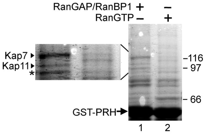
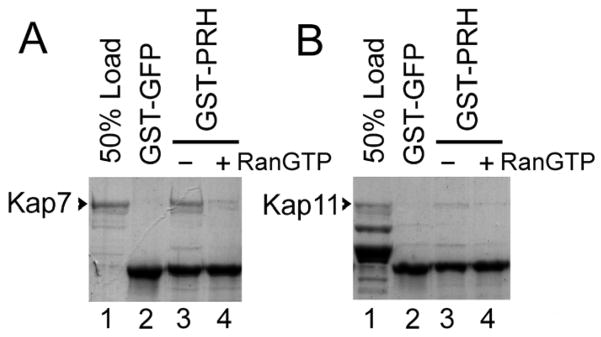
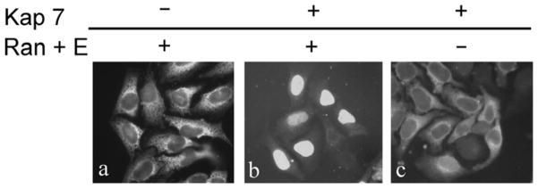
Proline-rich homeodomain (PRH)/hematopoietically expressed homeodomain (Hex) is a homeodomain protein that plays an important role in early embryonic patterning and hematopoiesis. PRH can act as either a tumor suppressor or an oncogene and its expression is dysregulated in certain types of lymphoid and myeloid leukemias. Aberrant exclusion of PRH from the nuclei has been associated with thyroid and breast cancers and a subset of myeloid leukemias. Accordingly, nuclear localization of PRH was found to be necessary for the inhibition of eIF4E-dependent transformation. Since PRH's nuclear-cytoplasmic localization has been associated with neoplastic transformation we sought to better understand how PRH is transported to the nuclear compartment. Here, we report an essential element that controls the mechanism of PRH nucleocytoplasmic trafficking, namely that it is imported into the nuclei by Karyopherin/Importin 7. Kap7 was identified as a binding partner for PRH in a GST-pull down from a HeLa cell protein lysate, followed by mass-spectrometry. The Kap7-PRH complex is dissociated in the presence of RanGTP, as expected for a nuclear import complex. Kap7 can bind directly to PRH in a GST-pull down assay with purified proteins, as well as mediates the transport of PRH to the nuclear compartment in a digitonin permeabilized cells assay. Finally, in vivo depletion of Kap7 dramatically reduces accumulation of PRH in the nucleus. Our data open the way for investigations of the mechanism of perturbed PRH localization in tumors and possible therapeutic interventions.


Figures

Figure 1
Figure 1. Kap7 interacts with PRH
a. Kap7 binds to GST-PRH in a GST-pull down assay from a total protein HeLa cell lysate. GST-PRH was expressed and purified from E. coli, and ~15 μg of GST-PRH was immobilized on glutathione-Sepharose and mixed with total HeLa cell lysate (lanes 1 and 2). Recombinant purified RanBP1 and RanGAP were added to the lysate (lane 1), to hydrolyze endogenous RanGTP. As a control, recombinant purified RanGTP was added to the lysate (lane 2) to promote dissociation of import Kap-cargo complexes. The proteins retained on the beads were analyzed by SDS-PAGE followed by Coomassie staining. A major protein band in the range 95–125 kDa (a characteristic for the Kap β family), was detected and analyzed by mass-spectrometry to be the karyopherin, Kap7. The left side insert (lanes 1a and 2a) is an enlargement of the 95–125 kDa region of the gel. b. Kap7 peptides identified by mass-spectrometry are depicted with highly significant expect values. c. Purified recombinant Kap7 binds directly to GST-PRH in a GST-pull down assay. PRH was expressed as a GST fusion in E. coli, purified and immobilized on glutathione-Sepharose. 1 μg of purified recombinant Kap7 was mixed with 1–2 μg of immobilized GST-PRH. After washing, the proteins retained on the beads were analyzed by SDS-PAGE followed by Coomassie staining. Lane 1 shows 50% of the Kap7 that was used in lanes 2–4. The addition of RanGTP reduced binding of Kap7 to GST-PRH (lane 4) as compared to lane 3, where no RanGTP was added. No binding of Kap7 to the control GST-GFP is detected (lane 2).
Figure 2
Figure 2. Kap7 mediates the import of PRH in digitonin permeabilized cells in a Ran and energy dependent manner
HeLa cells were permeabilized with digitonin, incubated for 25 minutes with the import mixtures, washed and fixed. Only the cargo imported into the nuclei is retained after washing. GST-GFP-PRH is imported into the nuclei in the presence of Kap7, Ran, RanBP1, RanGAP and NTF2 and an energy regenerating system (panel ii), but it is not imported in the absence of Kap7 (i) or in the absence of Ran and the energy regenerating system (iii). As expected, Kap7 cannot mediate the transport of the GFP tagged SV40 NLS to the nuclear compartment (panels iv, v, vi). Similar experiments were performed using Kapβ1 and Kapα2, which can mediate the transport of the SV40 NLS to the nuclear compartment in a Ran and energy dependent manner (panels viii). In the presence or absence of Ran and the energy regenerating system, Kapβ1 and Kapα2 did not mediate significant transport of GST-GFP-PRH to the nuclear compartment (Fig. 2x, xi, xii).
Figure 3
Figure 3. In vivo gene knockdown of Kap7 prevents accumulation of PRH in the nucleus
a. Hek 293 cells were co-transfected with a plasmid coding for Xpress-tagged PRH along with either siRNAs targeting Kap7 (panel a, i and ii) or Kapβ1 (panel a, v and vi). Additionally Hek 293 cells were co-transfected with a plasmid coding for GFP-NLS along with either siRNAs targeting Kap7 (panel a, iii and iv) or Kapβ1 (panel a, vii and viii). 96 hours post transfection, cells were washed, fixed, and stained with DAPI (Note that the DAPI stained cells depicted in panels a, i, iii, v, and vii are the same cells as in a, ii, iv, vi, viii, respectively). Immunofluorescence for Xpress-tagged PRH is shown in panels a, ii and vi. Depletion of Kap7 is associated with a striking decrease in the nuclear accumulation of PRH (panel a, ii) as compared to cells transfected with siRNA targeted to deplete Kapβ1 (panel a, vi). Fluorescence for GFP-NLS is shown in panels a, iv and viii. Depletion of Kap7 had no effect on the nuclear accumulation of GFP-NLS (panel a, iv). In contrast, cells transfected with siRNA targeted to deplete Kapβ1 (panel a, viii) exhibited reduced nuclear accumulation of GFP-NLS and a noticeable increase in cytoplasmic accumulation. b. Hep G2 cells were transfected with siRNAs targeted to Kap7 or Kapβ1. 96 hours post transfection, cells were washed, fixed and immunofluorescence for endogenous PRH was performed. Depletion of Kap7 is associated with a striking decrease in the nuclear accumulation of PRH (panel b, ii) as compared to control cells that were not transfected with siRNA (Mock) (panel b, i), or cells transfected with siRNA targeted to deplete Kapβ1 (panel b, iii). c. Quantitative RT-PCR utilizing gene specific DNA primers for Kap7 and Kapβ1 at 72 hours post siRNA transfection. Kap7 mRNA was significantly reduced in cells that were transfected with Kap7 siRNA as compared to a mock transfection, or cells transfected with Kapβ1 siRNA (n = 3, *p<0.005 relative to the mock transfection group). Additionally cells transfected with Kapβ1 siRNA, exhibited a significant reduction of Kapβ1 mRNA as compared to a mock transfection or cells transfected with Kap7 siRNA (n = 3, *p<0.005 relative to the mock transfection group). Error bars represent standard error of the mean (SEM). d. Western blotting using protein specific antibodies confirmed a significant reduction of Kap7 protein in cells that were transfected with Kap7 siRNA, but not cells transfected with Kapβ1 siRNA, or a mock transfection (n = 3, *p<0.005 relative to the mock transfection group). Additionally cells transfected with Kapβ1 siRNA, did not show a reduction of Kap7 protein, but did show a significant reduction of Kapβ1 protein (n = 3, *p<0.005 relative to the mock transfection group). Error bars represent SEM. e. Representative images for western blot data for experiment in c. Rows represent western blot data using respective antibodies against Kapβ1, Kap7 and GAPDH. All samples were normalized against the GAPDH loading control. Columns represent samples treated with siRNAs for Kapβ1 (β1), Kap7 (7), or the mock transfection (M).
Figure 4
Figure 4. Leptomycin B induces nuclear accumulation of PRH
K562 cells in culture were treated with leptomycin B (upper panels) or vehicle only (lower panels). After treatment the cells were fixed and PRH was detected using anti PRH antibodies followed by FITC-labeled secondary antibodies; left-side panels: confocal fluorescence images; middle panels: phase-contrast; right-side: overlay of the phase-contrast and PRH signal. The cytoplasmic areas, visible as rings in the phase-contrast images, are devoid of PRH in the leptomycin-treated cells (upper right panel), but not in the control cells (lower right).
Figure 5
Figure 5. The NLS recognized in PRH by Kap7 most likely includes the basic residues at the C-terminal of the homeodomain
A. PRH and Caudal, which share similar import pathways via Kap7, share high similarity within the domain responsible for binding to Kap7. The homeodomain regions of PRH and Caudal (dashed lines) are aligned. The thick bars represent α helical regions. Residues highlighted in gray are similar between PRH and Caudal. The underlined region of Caudal confers binding to Kap7 (48). Conserved basic residues within this region are marked by dark gray.

Similar articles

Cited by

References

    1. Crompton MR, Bartlett TJ, MacGregor AD, et al. Identification of a novel vertebrate homeobox gene expressed in haematopoietic cells. Nucleic Acids Res. 1992;20:5661–5667. - PMC - PubMed
    1. Martinez Barbera JP, Clements M, Thomas P, et al. The homeobox gene Hex is required in definitive endodermal tissues for normal forebrain, liver and thyroid formation. Development. 2000;127:2433–2445. - PubMed
    1. Manfioletti G, Gattei V, Buratti E, et al. Differential expression of a novel proline-rich homeobox gene (Prh) in human hematolymphopoietic cells. Blood. 1995;85:1237–1245. - PubMed
    1. Jayaraman PS, Frampton J, Goodwin G. The homeodomain protein PRH influences the differentiation of haematopoietic cells. Leuk Res. 2000;24:1023–1031. - PubMed
    1. Hromas R, Radich J, Collins S. PCR cloning of an orphan homeobox gene (PRH) preferentially expressed in myeloid and liver cells. Biochem Biophys Res Commun. 1993;195:976–983. - PubMed

Publication types

MeSH terms