Review
. 2009 Dec;31(4):305-25.
doi: 10.1007/s11357-009-9103-6.

Tissue elasticity and the ageing elastic fibre

Affiliations
Review

Tissue elasticity and the ageing elastic fibre

Michael J Sherratt. Age (Dordr). 2009 Dec.

Abstract

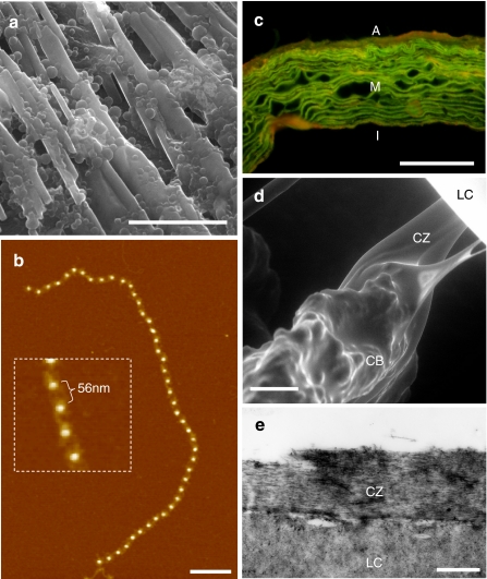
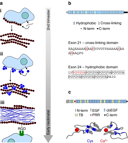
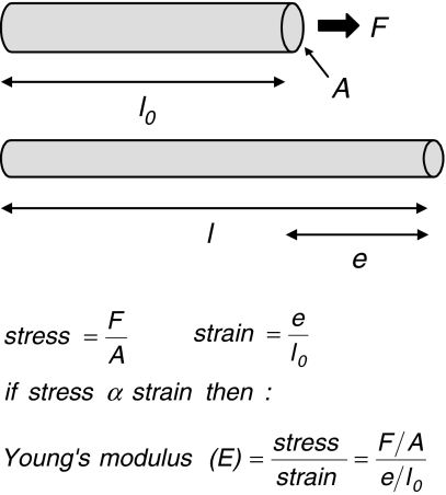
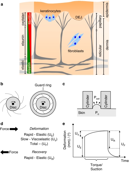
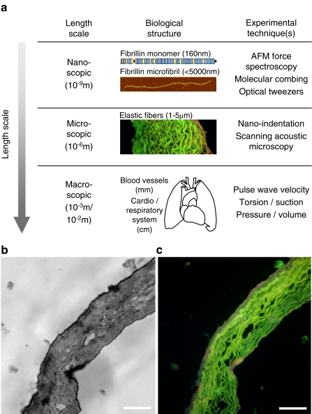
The ability of elastic tissues to deform under physiological forces and to subsequently release stored energy to drive passive recoil is vital to the function of many dynamic tissues. Within vertebrates, elastic fibres allow arteries and lungs to expand and contract, thus controlling variations in blood pressure and returning the pulmonary system to a resting state. Elastic fibres are composite structures composed of a cross-linked elastin core and an outer layer of fibrillin microfibrils. These two components perform distinct roles; elastin stores energy and drives passive recoil, whilst fibrillin microfibrils direct elastogenesis, mediate cell signalling, maintain tissue homeostasis via TGFβ sequestration and potentially act to reinforce the elastic fibre. In many tissues reduced elasticity, as a result of compromised elastic fibre function, becomes increasingly prevalent with age and contributes significantly to the burden of human morbidity and mortality. This review considers how the unique molecular structure, tissue distribution and longevity of elastic fibres pre-disposes these abundant extracellular matrix structures to the accumulation of damage in ageing dermal, pulmonary and vascular tissues. As compromised elasticity is a common feature of ageing dynamic tissues, the development of strategies to prevent, limit or reverse this loss of function will play a key role in reducing age-related morbidity and mortality.


Figures

Fig. 1a–e
Fig. 1a–e
Elastic fibre tissue distribution and composition. a, b The major elastic fibre components: elastin (a) and fibrillin microfibrils (b). a Environmental scanning electron microscopy (ESEM) image of the linear arrays and globules formed by coacervated recombinant tropoelastin. b AFM height image and high-resolution inset of a fibrillin microfibril isolated from young human skin (27-year-old male). The bright beads are raised 7–8 nm above the mica surface and are spaced 56 nm apart. c-e Composite elastic fibres are abundantly distributed in major arteries (c), whilst fibrillin microfibrils alone transmit forces between the ciliary muscle and the lens in the eye (d, e). c Fluorescence microscope image of haemotoxylin and eosin-stained ferret aorta. The green auto-fluorescence of elastic fibres, which are arranged into concentric lamellae in the medial (M) layer, is considerably enhanced by prior haemotoxylin and eosin staining (deCarvalho and Taboga 1996). The outer advential and inner intimal layers are indicated by A and I, respectively. d, e ESEM (d) and TEM (e) images of human ciliary zonules (CZ), which originate in the ciliary body (CB), intercalating with the lens capsule (LC) holding the lens in dynamic suspension. Scale bar 20 μm (a), 200 nm (b), 50 μm (c, d) and 500 nm (e)
Fig. 2a–c
Fig. 2a–c
Elastic fibre composition and assembly. a Assembly of fibrillin into microfibrils and the association of microfibrils with tropoelastin to form elastic fibres is a highly organised process which is limited to foetal and early neonatal development. i Secreted profibrillin is processed and assembled into pericellular microfibrils and microfibril bundles. ii Elastin globules which have assembled at the cell surface coalesce on the microfibril scaffold. iii In the core of the mature elastic fibre, ultrastructural analyses reveal twisted rope-like structures of highly cross-linked elastin (Ronchetti et al. 1998). Fibrillin microfibrils are mostly located at the microfibril periphery, where they can interact with cellular integrins via an RGD site on fibrillin-1 (Bax et al. 2003). b The structure of tropoelastin consists of alternating hydrophobic and cross-linking domains. Exon 21, for example, encodes a cross-linking domain in which pairs of lysine residues (K) are separated by two or three alanine (A) residues. In contrast, hydrophobic domains are characterised by repeating PGVGVA motifs (Keeley et al. 2002). c Fibrillin-1 is large (~320 kDa) modular glycoprotein, which, in addition to unique N- and C-terminal regions (N-term and C-term) and a potentially flexible proline-rich region (PRR), is predominantly composed of repeating eight-cysteine (also known as TB modules) and EGF-like domains, which may (cbEGF) or may not (EGF) bind calcium. The cbEGF domains play a major role in maintaining fibrillin microfibril structure. Each domain is stabilised by three cys-cys disulphide bonds (indicated in blue on the ribbon model of two contiguous fibrillin-1 cbEGF domains) and by a single bound Ca2+ (Downing et al. ; Wess et al. 1998)
Fig. 3a, b
Fig. 3a, b
Quantifying elasticity. a The elastic modulus defines the degree to which a material deforms when a tensile force is applied. Where a rod of length l0 and cross-sectional area A is stretched to a length l by a force F, the elastic modulus (E) is calculated from the stress divided by the strain. b The value of E relates to the biological function of a macromolecules; the elastic modulus of fibrillar collagen (1,200 MPa), for example, is relatively high reflecting the role of collagen fibrils in resisting tensile forces (Gosline et al. 2002), in contrast E for elastin is low (1.1 MPa) (Aaron and Gosline 1981) and a small force will produce a large extension. The elastic modulus of fibrillin microfibrils, however, remains controversial with estimates ranging from 1.0 MPa (Aaron and Gosline 1981) to 96 MPa (Sherratt et al. 2003)
Fig. 4a–e
Fig. 4a–e
Structure and mechanical function of the skin. a The elastic fibre system in skin is confined to the dermis where composite elastic fibres in the reticular dermis give way to arrays of fibrillin microfibril bundles at the DEJ. The fibrillin microfibrils of the elastic fibre system are synthesised by both keratinocytes and dermal fibroblasts. b, c Methods for testing the mechanical properties of skin include: torsion, induced by a rotating disk (b), where the area of skin may be restrained with a guard ring and suction (c), where the lowered external air pressure (Pext) in a cylinder causes skin deformation due to the internal pressure of the tissue (Po). d, e Both methods induce a total deformation (UF) which is the result of an initial rapid (UE) and a slower viscoelastic (UV) deformation. Release of the torque or suction is followed by a rapid recovery (UR)
Fig. 5a–c
Fig. 5a–c
Measuring biomechanical properties at increasing length scales. a In general, the elastic moduli of the constituent ECM molecules are higher than elastic moduli of the connective tissues themselves (Akhtar et al. 2009). In order, therefore, to understand the mechanical role played by age-related changes in molecular abundance, distribution and structure it is necessary to characterise the mechanical properties of tissues at the nano-scopic, micro-scopic and macro-scopic lengths scales. b, c Scanning acoustic microscopy (SAM) image of an unfixed cryo-sectioned ferret aorta (b) and a fluorescence microscopy image of the same section post-stained with haematoxylin and eosin (c). Variations in wavespeed in the SAM image (which are correlated with material stiffness) closely match the distribution of elastic fibres imaged in the fluorescence microscope. Scale bar 100 μm

References

    1. Aaron BB, Gosline JM. Elastin as a random-network elastomer: a mechanical and optical analysis of single elastin fibers. Biopolymers. 1981;20:1247–1260.
    1. Agache PG, Monneur C, Leveque JL, Derigal J. Mechanical-properties and youngs modulus of human-skin in vivo. Arch Dermatol Res. 1980;269:221–232. - PubMed
    1. Akhtar R, Schwarzer N, Sherratt MJ, Watson REB, Graham HK, Trafford AW, Mummery PM, Derby B (2009) Nanoindentation of histological specimens: mapping the elastic properties of soft tissues. J Mater Res 24:638–646 - PMC - PubMed
    1. Andreotti L, Bussotti A, Cammelli D, Aiello E, Sampognaro S. Connective-tissue in aging lung. Gerontology. 1983;29:377–387. - PubMed
    1. Antoniou J, Steffen T, Nelson F, Winterbottom N, Hollander AP, Poole RA, Aebi M, Alini M. The human lumbar intervertebral disc - Evidence for changes in the biosynthesis and denaturation of the extracellular matrix with growth, maturation, ageing, and degeneration. J Clin Invest. 1996;98:996–1003. - PMC - PubMed

Publication types