. 2009 Aug 4;48(30):7160-8.
doi: 10.1021/bi900830g.

The flexibility of a distant loop modulates active site motion and product release in ribonuclease A

Affiliations

The flexibility of a distant loop modulates active site motion and product release in ribonuclease A

Nicolas Doucet et al. Biochemistry. .

Abstract

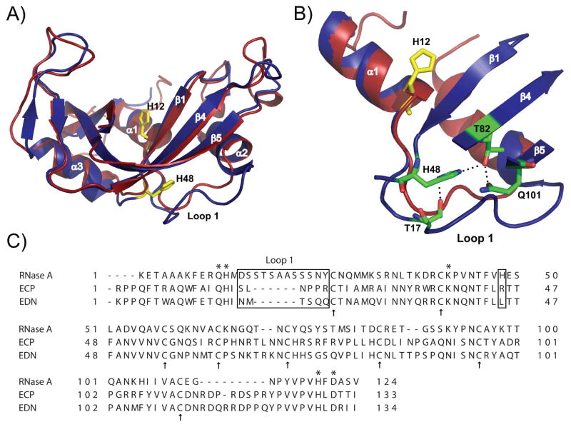
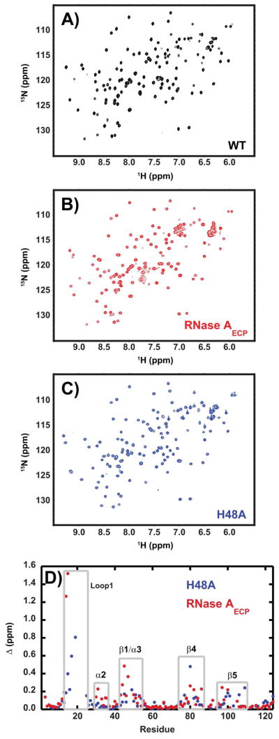
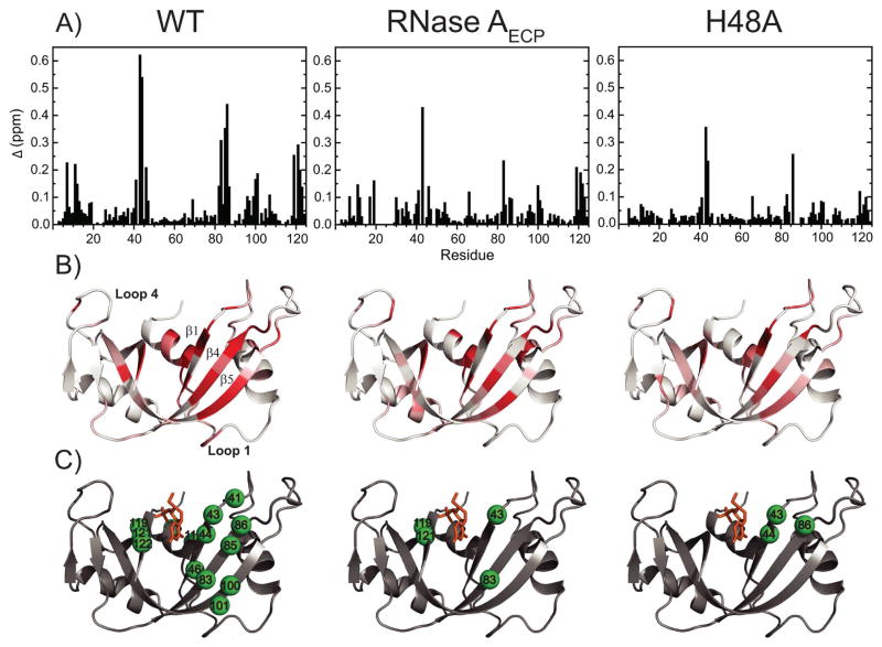
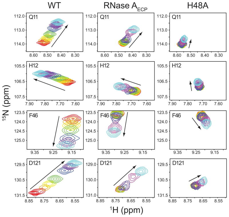
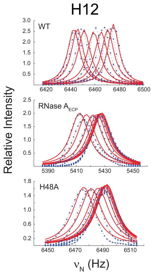
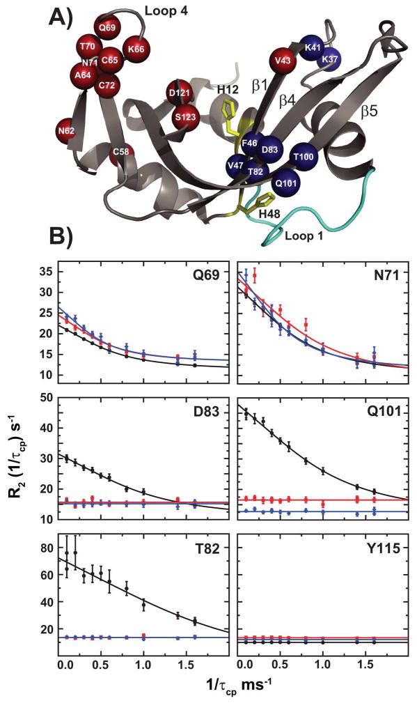
The role of the flexible loop 1 in protein conformational motion and in the dissociation of enzymatic product from ribonuclease A (RNase A) was investigated by creation of a chimeric enzyme in which a 6-residue loop 1 from the RNase A homologue, eosinophil cationic protein (ECP), replaced the 12-residue loop 1 in RNase A. The chimera (RNase A(ECP)) experiences only local perturbations in NMR backbone chemical shifts compared to WT RNase A. Many of the flexible residues that were previously identified in WT as involved in an important conformational change now experience no NMR-detected millisecond motions in the chimera. Likewise, binding of the product analogue, 3'-CMP, to RNase A(ECP) results in only minor chemical shift changes in the enzyme similar to what is observed for the H48A mutant of RNase A and in contrast to WT enzyme. For both RNase A(ECP) and H48A there is a 10-fold decrease in the product release rate constant, k(off), compared to WT, in agreement with previous studies indicating the importance of flexibility in RNase A in the overall rate-limiting product release step. Together, these NMR and biochemical experiments provide additional insight into the mechanism of millisecond motions in the RNase A catalytic cycle.


Figures

Figure 1
Figure 1
Structural comparison of bovine ribonuclease A (RNase A) and human Eosinophil Cationic Protein (ECP). A) Superposition of RNase A (blue structure, PDB code 1FS3 (35)) and ECP (red structure, PDB code 1DYT (33)). H12 highlights the position of the active site relative to the position of His48 and loop 1 in RNase A. B) Zoomed view of a few atomic interactions between His48 and loop 1 in RNase A. Note the longer loop 1 in RNase A (D14SSTSAASSSNY25) relative to the shorter loop 1 of ECP (S17LNPPR22, ECP numbering). C) Primary sequence alignment of RNase A, ECP and EDN. Active site positions Gln11, His12, Lys41, His119 and Asp121 are marked with stars while cysteine residues forming the strictly conserved four disulfide bridges are represented by arrows. Loop 1 and position 48 are boxed. Alignment was performed with TCoffee Expresso (36) using PDB coordinates 7RSA (32), 1DYT (37) and 1GQV (38).
Figure 2
Figure 2
Chemical shift changes caused by mutation. 1H-15N HSQC spectra for (A) wild-type, (B) RNase AECP, and (C) H48A enzymes. The spectra were acquired at 14.1 T and 298K, pH = 6.4. In (D) chemical shift differences for WT vs. H48A (blue) and WT vs. RNase AECP are shown as a function of amino acid sequence. Gray boxed areas depict protein regions with larger than average chemical shift disturbances. (Δ (ppm) = [(Δδ2HN + Δδ2 N/25)/2]1/2) in which δ is the difference in chemical shift between the two proteins for NH and HN nuclei (52).
Figure 3
Figure 3
1H-15N chemical shift differences upon 3′-CMP binding to WT RNase A, RNase AECP, and H48A. (A) 1H-15N chemical shift differences Δ (ppm) mapped on the primary sequence of WT, RNase AECP, and H48A. The value of Δ was calculated between a ligand-free 1H-15N HSQC (mole ratio, 3′-CMP/Enzyme = 0) and a 3′-CMP-bound 1H-15N HSQC (mole ratio, 3′-CMP/Enzyme = 12) according to the following equation: (Δ (ppm) = [(Δδ2 HN + Δδ2 N/25)/2]1/2) (52). (B) Mapping of Δ (ppm) on the three-dimensional structure of RNase A (PDB code 1RPF (51)). The white to red gradient corresponds to Δ values ranging from 0 ppm (white) to > 0.15 ppm (dark red). (C) Same view as in (B) showing residues with Δ > 0.15 ppm as green spheres. 3′-CMP is shown as orange sticks representation.
Figure 4
Figure 4
Kinetics of 3′-CMP binding to WT RNase A, RNase AECP and mutant H48A. Titration of 3′-CMP is followed by monitoring the changes in backbone chemical shifts observed in 1H-15N-HSQC spectra for WT RNase A, RNase AECP and mutant H48A. Titration points are shown for residues Gln11, His12, Phe46 and Asp121 for [3′-CMP]:[RNase A] mole ratios of 0 (red) 0.174 (orange), 0.393 (yellow), 0.691 (green), 1.31 (blue), 2.71 (purple), 6 (magenta), and 12 (cyan). Arrows indicate the direction of resonance shift with increasing [3′-CMP]. The magnitude of 3′-CMP binding-induced chemical shifts is smaller in mutant H48A than in RNase AECP.
Figure 5
Figure 5
3′-CMP titration of RNase A. The titration curves of residue His12 are shown for WT, RNase AECP and H48A. Fitted line shapes (red) are superimposed on the experimental NMR data (blue).
Figure 6
Figure 6
Conformational dynamics in RNase A. (A) Residues in RNase AECP undergoing conformational exchange with a ΔR2 (1/τcp) greater than 1.5 s−1 are plotted on the structure of RNase A (PDB code 7RSA (32)). Red spheres = similar rate constants (kex) in WT and RNase AECP; Blue spheres = Decreased kex values in RNase AECP relative to WT. H12 and H48 are shown as yellow sticks and loop 1 is shown in cyan. B) CPMG dispersion curves for WT RNase A (black circles), RNase AECP (red squares) and mutant H48A (blue diamonds). 15N relaxation dispersion curves are shown for two residues displaying similar conformational exchange in the three enzymes (Gln69 and Asn71), and three residues displaying significantly affected kex in both RNase AECP and mutant H48A (Thr82, Asp83 and Gln101). Y115 is represented as a motionless control. Fitted lines to data points are from single-field fits at 14.1 T using equation (1).

References

    1. Daniel RM, Dunn RV, Finney JL, Smith JC. The role of dynamics in enzyme activity. Annu Rev Biophys Biomol Struct. 2003;32:69–92. - PubMed
    1. Eisenmesser EZ, Millet O, Labeikovsky W, Korzhnev DM, Wolf-Watz M, Bosco DA, Skalicky JJ, Kay LE, Kern D. Intrinsic dynamics of an enzyme underlies catalysis. Nature. 2005;438:117–121. - PubMed
    1. Kovrigin EL, Loria JP. Enzyme dynamics along the reaction coordinate: critical role of a conserved residue. Biochemistry. 2006;45:2636–2647. - PubMed
    1. Boehr DD, McElheny D, Dyson HJ, Wright PE. The dynamic energy landscape of dihydrofolate reductase catalysis. Science. 2006;313:1638–1642. - PubMed
    1. Blacklow SC, Raines RT, Lim WA, Zamore PD, Knowles JR. Triosephosphate isomerase catalysis is diffusion controlled. Biochemistry. 1988;27:1158–1167. - PubMed

Publication types

MeSH terms

Substances