. 2009 Jul 9:6:100.
doi: 10.1186/1743-422X-6-100.

Experimental induction of proventricular dilatation disease in cockatiels (Nymphicus hollandicus) inoculated with brain homogenates containing avian bornavirus 4

Affiliations

Experimental induction of proventricular dilatation disease in cockatiels (Nymphicus hollandicus) inoculated with brain homogenates containing avian bornavirus 4

Ady Y Gancz et al. Virol J. .

Abstract

Background: Proventricular dilatation disease (PDD) is a fatal disorder of psittacine birds worldwide. The disease is characterized by lymphoplasmacytic infiltration of the central and peripheral nervous systems, leading to gastrointestinal motility and/or central nervous system dysfunction. Recently, we detected a significant association between avian bornavirus (ABV) infection and clinical signs of PDD in psittacines. However, it remains unclear whether ABV infection actually causes PDD. To address this question, we examined the impact of ABV inoculation on the cockatiel (Nymphicus hollandicus).

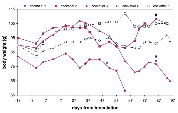
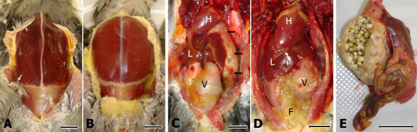
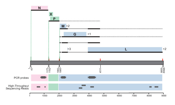
Results: Five cockatiels were inoculated via multiple routes (intramuscular, intraocular, intranasal, and oral) with a brain homogenate derived from either a PDD(+) avian bornavirus 4 (ABV4) (+) case (n = 3 inoculees) or from a PDD(-) ABV(-) control (n = 2 inoculees). The control birds remained free of clinical or pathological signs of PDD, and tested ABV(-) by RT-PCR and immunohistochemistry (IHC). In contrast, all three cockatiels inoculated with ABV4(+) brain homogenate developed gross and microscopic PDD lesions, and two exhibited overt clinical signs. In numerous tissues, ABV RT-PCR and sequence analysis demonstrated the presence of ABV4 RNA nearly identical to that in the inoculum. ABV was detected in the central nervous system of the three ABV-inoculees by IHC. Pyrosequencing to investigate the viral flora in the ABV4(+) inoculum uncovered 7 unique reads sharing 73-100% nucleotide sequence identity with previously identified ABV sequences and 24 reads sharing 40-89% amino acid sequence identity with viruses in the Retroviridae and Astroviridae families. Of these candidate viral species, only ABV RNA was recovered from tissues of the inoculated birds.

Conclusion: In this study, the clinical and pathological manifestations of PDD were induced by inoculation of cockatiels with brain homogenates containing avian bornavirus 4. By using high throughput pyrosequencing an in-depth view of the viral content of the inoculum was achieved, revealing that of 3 candidate virus families detected, only the presence of ABV RNA correlated with the development of PDD. This study provides evidence of a causal association between ABV4 infection and PDD in cockatiels.


Figures

Figure 1
Figure 1
Body weights of the ABV4-inoculated and sham-inoculated cockatiels during the study period. The ABV4-inoculated birds (cockatiels 1–3), are shown in bald lines with solid symbols. Cockatiels 4,5 are the control birds; "*" marks the first observation of undigested seeds in the feces; "#" marks the first detection of ABV4 RNA in a choanal or cloacal swab by RT-PCR. Note the continuous decrease in BW of cockatiels 1 and 3 starting on day 21 PI and 31 PI, respectively.
Figure 2
Figure 2
Macroscopic pathological findings in an ABV4-inoculated cockatiel. Bar size = 1 cm; F = peritoneal fat; H = heart; L = right liver lobe; V = ventriculus; A) Markedly reduced pectoral muscle mass and subcutaneous fat stores are clearly seen in an ABV4-inoculated cockatiel (cockatiel 3). B) Normal pectoral muscle mass and subcutaneous fat stores in a control bird (cockatiel 5). C) The peritoneal cavity of cockatiel 3, showing a severely distended and thin-walled proventriculus (arrows) that is visible well beyond the left liver lobe. The ventriculus is mildly distended and the peritoneal fat is dramatically reduced. D) In cockatiel 5, the proventriculus is of normal size, and; therefore, completely hidden behind the left liver lobe. Note the abundant peritoneal fat stores. E) The proventriculus and intestine of cockatiel 3. The thin wall has been cut, exposing a large amount of undigested seeds. Whole seeds are also visible through the intestinal wall.
Figure 3
Figure 3
Histological and immunohistochemical findings in ABV4-inoculated cockatiels. A) Myenteric ganglioneuritis (arrow) in the crop of cockatiel 2 (hematoxyline and eosin [H&E] staining); B) Myenteric ganglioneuritis (arrow) in the ventriculus of cockatiel 3 (H&E staining). C) Positive IHC staining for ABVN associated with focal gliosis (arrow) in the cerebrum of cockatiel 1. Here, the staining appears to be mainly extracellular. D) For negative control, a section parallel to that in C was stained, using pre-immune rabbit serum instead of the anti-ABVN antibody. E) Widespread positive IHC staining for ABVN of neurons and glial cells of the cerebrum of cockatiel 2. At greater magnification, staining of a large neuron and its dendrites is shown (inset). F) For negative control, a section parallel to that in E was stained, using pre-immune rabbit serum instead of the anti-ABVN antibody.
Figure 4
Figure 4
Transmission electron microscopy images. A) Brain homogenate from an ABV4(+) PDD(+) African grey parrot (the inoculum used in this study). Three spherical virus-like particles approximately 60 nm in diameter are shown [arrows] (negative staining with uranyl acetate). B) A virus-like particle from the same specimen in "A" shown at greater magnification. This particle is 98 nm in diameter (negative staining with uranyl acetate). C) A virus-like particle from the brain of cockatiel 1. This particle is 99 nm in diameter and is showing bold projections on its circumference (negative staining with uranyl acetate).
Figure 5
Figure 5
Recovery of sequences matching ABV from the inoculum by high throughput pyrosequencing and RT-PCR. The location of seven unique RNA sequences, recovered from the inoculum by high throughput pyrosequencing, is shown. These sequences share 73–100% sequence identity with existing ABV sequences (Additional file 2). In addition, the location of the RT-PCR primers for the N, M, and genes is shown, all of which yielded products that were consistent with ABV4 genome.

References

    1. Daoust PY, Julian RJ, Yason CV, Artsob H. Proventricular impaction associated with nonsuppurative encephalomyelitis and ganglioneuritis in two Canada geese. J Wildl Dis. 1991;27:513–517. - PubMed
    1. Doneley RJ, Miller RI, Fanning TE. Proventricular dilatation disease: an emerging exotic disease of parrots in Australia. Aust Vet J. 2007;85:119–123. doi: 10.1111/j.1751-0813.2007.00109.x. - DOI - PMC - PubMed
    1. Gregory C, Latimer KS, Niagro F, Ritchie BW, Campagnoli RP, Norton TM, Greenacre CB. A review of proventricular dilation syndrome. J Assoc Avian Vet. 1994;8:69–75.
    1. Sullivan ND, Mackie JT, Miller RI, Giles A. First case of psittacine proventricular dilatation syndrome (macaw wasting disease) in Australia. Aust Vet J. 1997;75:674. doi: 10.1111/j.1751-0813.1997.tb15371.x. - DOI - PubMed
    1. Berhane YSD, Newman S, Taylor M, Nagy E, Binnington B, Hunter B. Peripheral neuritis in psittacine birds with proventricular dilation disease. Avian Pathol. 2001;30:563–570. doi: 10.1080/03079450120078770. - DOI - PubMed

MeSH terms

LinkOut - more resources