. 2009 Sep 4;284(36):23912-24.
doi: 10.1074/jbc.M109.036483. Epub 2009 Jul 9.

Phosphatidylinositol 3-kinase/Akt pathway targets acetylation of Smad3 through Smad3/CREB-binding protein interaction: contribution to transforming growth factor beta1-induced Epstein-Barr virus reactivation

Affiliations

Phosphatidylinositol 3-kinase/Akt pathway targets acetylation of Smad3 through Smad3/CREB-binding protein interaction: contribution to transforming growth factor beta1-induced Epstein-Barr virus reactivation

Lassad Oussaief et al. J Biol Chem. .

Abstract

Epstein-Barr virus, a ubiquitous human herpesvirus, is associated with the development of carcinomas and lymphomas. We previously showed that transforming growth factor beta1 (TGF-beta1) mediated the virus to enter the lytic cycle, which is triggered by expression of Z Epstein-Barr virus replication activator (ZEBRA), through the ERK 1/2 MAPK signaling pathway. We report here that Akt, activated downstream from ERK 1/2, was required for TGF-beta1-induced ZEBRA expression and enabled Smad3, a mediator of TGF-beta1 signaling, to be acetylated by direct interaction with the co-activator CREB-binding protein and then to regulate TGF-beta1-induced ZEBRA expression.


Figures
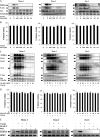
FIGURE 1.
FIGURE 1.
Inhibitors of PI3-K/Akt signaling pathway abolished TGF-β1-induced ZEBRA expression. A and C, Mutu-I, Kem-I, and Sav-I cells were treated with increasing concentration of wortmannin (Wort) (0.01, 0.05, 0.1, or 0.2 μm) (A) or Akt inhibitor X (Akt inh X) (0.1, 1, 2, 5, or 10 μm) (C) for 1 h, prior to incubation with TGF-β1 (2 ng/ml). Seventeen hours later, the cells were harvested and lysed. Equal amounts of protein were separated by SDS-PAGE and analyzed by Western blotting with antibodies to ZEBRA, phospho-Akt, Akt, and tubulin. B and D, viability assay was performed with the LIVE/DEAD reduced biohazard viability/cytotoxicity kit (Molecular Probes, Invitrogen). The ability of this kit to detect cell death was controlled using a treatment with 0.4 mm of MnCl2 (not shown). E, RT-PCR assay of ZEBRA was performed. 3 μg of total RNA from Mutu-I, Kem-I, and Sav-I cells pretreated, respectively, with 0.05, 0.05, and 0.1 μm of wortmannin or 10 μm of Akt inhibitor X for 1 h and then stimulated with TGF-β1 (2 ng/ml) for 17 h were reverse transcribed. cDNA coding for ZEBRA was then analyzed by PCR; cDNA of hypoxanthine-guanine phosphoribosyltransferase (HPRT) was used as an internal control.
FIGURE 2.
FIGURE 2.
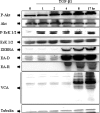
Time course of TGF-β1-induced phosphorylation of Akt and ERK 1/2, and EBV lytic gene expression in Mutu-I cells. Mutu-I cells were incubated in the presence of TGF-β1 (2 ng/ml) for various periods of time. The cells were lysed, and equal amounts of proteins were separated by SDS-PAGE. Phosphorylated Akt and Akt were analyzed, respectively, with anti-phospho-Akt and Akt antibodies by Western blotting. The membrane was then reprobed separately with a panel of specific antibodies directed against phospho-ERK 1/2, ERK 1/2, ZEBRA, EA-D, EA-R, and VCA. The amounts of protein loaded were assayed by reprobing the membrane with anti-tubulin antibody.
FIGURE 3.
FIGURE 3.
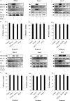
TGF-β1-induced Akt phosphorylation in an ERK 1/2-dependent manner. A, Mutu-I, Kem-I, and Sav-I cells were incubated in the presence of 20 μm of U0126 for 1 h and then treated by TGF-β1 (2 ng/ml) for 17 h. The cells were harvested, washed, and lysed. Equal amounts of protein were analyzed by Western blotting using specific antibodies against phospho-Akt, Akt, phospho-ERK 1/2, and ERK 1/2. The amounts of protein loaded were assayed by reprobing the membrane with anti-tubulin antibody. C, Mutu-I, Kem-I, and Sav-I cells were treated with 10 μm of Akt inhibitor X (Akt inh X), prior to TGF-β1 (2 ng/ml) stimulation. Seventeen hours later, the cells were harvested and resuspended in Laemmli sample buffer. Equals amounts of protein were separated by SDS-PAGE and analyzed by Western blotting using phospho-Akt, Akt, phospho-ERK 1/2, ERK 1/2, and tubulin antibodies. B and D, viability assay was performed with the LIVE/DEAD reduced biohazard viability/cytotoxicity kit (Molecular Probes, Invitrogen). The ability of this kit to detect cell death was controlled using a treatment with 0.4 mm of MnCl2 (not shown).
FIGURE 4.
FIGURE 4.
Time course of TGF-β-induced Smad3 phosphorylation and acetylation. A, Mutu-I cells were treated with TGF-β1 (2 ng/ml) for various periods of time. At the indicated time points, the cells were harvested, and lysed. Equal amounts of protein were separated by SDS-PAGE and analyzed by Western blotting with antibodies to phospho-Akt, Akt, phospho-Smad3, Smad3, ZEBRA, and tubulin. B, simultaneously, cell lysates were immunoprecipitated (IP) with anti-Smad3 antibody, and acetylated Smad3 was detected by immunoblot with anti-acetylated lysine antibody.
FIGURE 5.
FIGURE 5.
PI3-K/Akt pathway regulates TGF-β1-induced Smad3 acetylation without any effect on Smad3 phosphorylation, and translocation. A, cells were treated or not with 10 μm of Akt inhibitor X (Akt inh X) or 10 μm SB-431542 for 1 h and then simulated with TGF-β1 (2 ng/ml) for 6 h. The cells were harvested, washed, and resuspended in Laemmli sample buffer. Cell extracts were analyzed by Western blotting against phospho-Akt, Akt, phospho-Smad3, Smad3, ZEBRA, and tubulin. B, nuclear and cytosolic extracts were prepared as described under “Experimental Procedures.” Equal amounts of each extracts were fractioned by SDS-PAGE, and the content of Smad3 was determined by Western blotting using anti-Smad3 antibody. Loading of nuclear and cytosolic fractions was assayed by blotting with antibodies respectively to HP1G (for nuclear fraction) and tubulin (for cytosolic fraction). C, cell lysates were immunoprecipitated (IP) with anti-Smad3 antibody, and acetylated Smad3 was detected by immunoblot with anti-acetyllysine antibody.
FIGURE 6.
FIGURE 6.
Both Akt inhibitor X and wortmannin impair TGF-β-induced Smad3 association with CBP. Mutu-I, Kem-I, and Sav-I cells were treated, respectively, with 0.05, 0.05, and 0.1 μm of wortmannin (Wort) (A and B), 10 μm of Akt inhibitor X (Akt inh X) (C and D), or 10 μm of SB-431542 (A–D) for 1 h, prior to incubation with 2 ng/ml of TGF-β1. The cell lysates were immunoprecipitated (IP) with Smad3 (A and C) or CBP (B and D) antibodies, respectively. The immunoprecipitates were immunoblotted with anti-Smad3 or anti-CBP antibodies as indicated. The membranes were then reprobed with anti-acetyllysine antibody.
FIGURE 7.
FIGURE 7.
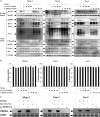
Lys-CoA-Tat inhibits TGF-β1-induced Smad3 acetylation and EBV-lytic cycle. A, Mutu-I, Kem-I, and Sav-I cells pretreated, respectively, with 10, 10, and 20 μm of Lys-CoA-Tat or 20 μm of Ac-DDDD-Tat for 1 h were stimulated with TGF-β1 (2 ng/ml) for 17 h. The cell lysates were immunoprecipitated (IP) with anti-Smad3 antibody, and acetylated Smad3 was detected by immunoblot with anti-acetyllysine antibody. Equal amounts of protein were separated by SDS-PAGE and analyzed by Western blotting with antibodies to ZEBRA, EA, VCA, phosphoSmad3, Smad3, phospho-Akt, Akt, and tubulin. B, viability assay was performed with the LIVE/DEAD reduced biohazard viability/cytotoxicity kit (Molecular Probes, Invitrogen). The ability of this kit to detect cell death was controlled using a treatment with 0.4 mm of MnCl2 (not shown). C, 3 μg of total RNA from Mutu-I, Kem-I, and Sav-I cells pretreated, respectively, with 10, 10, and 20 μm of Lys-CoA-Tat or 20 μm of Ac-DDDD-Tat for 1 h and then stimulated with TGF-β1 (2 ng/ml) for 17 h, were reverse transcribed. cDNA coding for ZEBRA was then analyzed by PCR; cDNA of hypoxanthine-guanine phosphoribosyltransferase (HPRT) was used as an internal control.
FIGURE 8.
FIGURE 8.
CBP shRNA inhibits TGF-β1-induced Smad3 acetylation and EBV-lytic cycle. A, Mutu-I, Kem-I, and Sav-I cells infected with CBP shRNA lentiviral particles or control particles were stimulated or not with TGF-β1 (2 ng/ml) for 17 h. The cell lysates were immunoprecipitated (IP) with anti-Smad3 antibody, and acetylated Smad3 was detected by immunoblot with anti-acetyllysine antibody. Simultaneously, equal amounts of protein from total lysate were separated by SDS-PAGE and analyzed by Western blotting with antibodies to ZEBRA, EA, VCA, phospho-Smad3, Smad3, phospho-Akt, Akt, and tubulin. B, 3 μg of total RNA from Mutu-I, Kem-I, and Sav-I cells infected with CBP shRNA lentiviral particles or control particles stimulated or not with TGF-β1 (2 ng/ml) for 17 h were reverse transcribed. cDNA coding for ZEBRA was then analyzed by PCR; cDNA of hypoxanthine-guanine phosphoribosyltransferase (HPRT) was used as an internal control.
FIGURE 9.
FIGURE 9.
Acetylation of Smad3 participates in TGF-β1-induced Zp activation. A, schematic representation of the Smad3 protein and its mutant used in transient transfection experiments. B, the DG75 cells were transiently transfected with reporter gene construct containing the wild type Zp (−234 to +12) inserted upstream of the CAT gene (Control), or with a combination of expression plasmids for wild type of Smad3 (Smad3 wt) or its mutant (Smad3 mut), CBP, or empty vectors (pcDNA3.1 and pCMV2N3T) as indicated. The positive control consisted of Zp (−234 to +12) transfected DG75 cells and treated by 20 ng/ml of phorbol 12-myristate 13-acetate (PMA) immediately following transfection. Twenty-four hours later, the cells were harvested and lysed, and CAT activity was quantified as described under “Experimental Procedures.” The error bars represent the standard deviation. Significant differences between control and the other samples were determined by Student's t test; *, p < 0.05. C, cell lysates were immunoprecipitated (IP) with anti-Smad3 antibody, and equal amounts of immunoprecipitated Smad3 were analyzed by Western blotting with anti-acetyllysine antibodies.
FIGURE 10.
FIGURE 10.
Proposed mechanism of TGF-β1-mediated ZEBRA expression. Canonical (implicating Smad3) as well as noncanonical (implicating PI3-K/Akt) pathways, in concert, contribute to TGF-β1-mediated ZEBRA expression. TGF-β1 mediates its activity through binding to its receptor II which recruits and phosphorylates its receptor I/ALK5; Smad3 phosphorylation takes place and translocation to the nucleus occurs. However, activated Akt, the non-Smad mediator of TGF-β1 directed CBP-Smad3 interaction and Smad3 acetylation, which enables the increase of ZEBRA expression.

Similar articles

Cited by

References

    1. Desgranges C., de-The G., Wolf H., zur Hausen H. (1975) IARC Sci. Publ. 11, 191–193 - PubMed
    1. Henle W., Henle G., Niederman J. C., Klemola E., Haltia K. (1971) J. Infect. Dis. 124, 58–67 - PubMed
    1. Pallesen G., Hamilton-Dutoit S. J., Rowe M., Young L. S. (1991) Lancet 337, 320–322 - PubMed
    1. Kieff (2007) Epstein-Barr Virus and Its Replication, Lippincott William & Wilkins, Philadelphia, PA
    1. Ragoczy T., Miller G. (1999) J. Virol. 73, 9858–9866 - PMC - PubMed

Publication types

MeSH terms

LinkOut - more resources