. 2009 Oct;155(Pt 10):3247-3259.
doi: 10.1099/mic.0.029124-0. Epub 2009 Jul 9.

Deletion of Braun lipoprotein gene (lpp) and curing of plasmid pPCP1 dramatically alter the virulence of Yersinia pestis CO92 in a mouse model of pneumonic plague

Affiliations

Deletion of Braun lipoprotein gene (lpp) and curing of plasmid pPCP1 dramatically alter the virulence of Yersinia pestis CO92 in a mouse model of pneumonic plague

Stacy L Agar et al. Microbiology (Reading). 2009 Oct.

Abstract

Deletion of the murein (Braun) lipoprotein gene, lpp, attenuates the Yersinia pestis CO92 strain in mouse models of bubonic and pneumonic plague. In this report, we characterized the virulence of strains from which the plasminogen activating protease (pla)-encoding pPCP1 plasmid was cured from either the wild-type (WT) or the Deltalpp mutant strain of Y. pestis CO92 in the mouse model of pneumonic infection. We noted a significantly increased survival rate in mice infected with the Y. pestis pPCP(-)/Deltalpp mutant strain up to a dose of 5000 LD(50). Additionally, mice challenged with the pPCP(-)/Deltalpp strain had substantially less tissue injury and a strong decrease in the levels of most cytokines and chemokines in tissue homogenates and sera when compared with the WT-infected group. Importantly, the Y. pestis pPCP(-)/Deltalpp mutant strain was detectable in high numbers in the livers and spleens of some of the infected mice. In the lungs of pPCP(-)/Deltalpp mutant-challenged animals, however, bacterial numbers dropped at 48 h after infection when compared with tissue homogenates from 1 h post-infection. Similarly, we noted that this mutant was unable to survive within murine macrophages in an in vitro assay, whereas survivability of the pPCP(-) mutant within the macrophage environment was similar to that of the WT. Taken together, our data indicated that a significant and possibly synergistic attenuation in bacterial virulence occurred in a mouse model of pneumonic plague when both the lpp gene and the virulence plasmid pPCP1 encoding the pla gene were deleted from Y. pestis.


Figures

Fig. 1.
Fig. 1.
Survival analysis of mice infected with WT and mutant Y. pestis strains. Following i.n. inoculation with various doses of WT, Δlpp, pPCP and pPCPlpp mutants of Y. pestis, animals were monitored for morbidity and mortality. Those infected with a 250 LD50 dose of either WT (•) or Δlpp (○) Y. pestis died by days 3 and 5, respectively. However, mice infected with either a 250 LD50 (▵) or a 2500 LD50 (□) dose of the pPCPlpp strain showed a 100 % survival rate, while those infected with the pPCP strain showed 30 % (▾) and 20 % (▪) survival rates, respectively. At the highest dose tested, 5000 LD50, 50 % of the mice infected with the pPCPlpp strain survived (◊), while no mice infected with the pPCP strain survived (⧫). By Kaplan–Meier analysis, only the group infected with a 5000 LD50 dose of the pPCP strain was not significantly different from the WT- and Δlpp-infected groups. All other treatment groups were significantly different (P<0.001).
Fig. 2.
Fig. 2.
Histopathology of mouse tissues following infection with WT and mutant strains. Groups of five mice were infected with WT Y. pestis or Δlpp, pPCP or pPCPlpp mutant strains and euthanized at 1 and 48 h p.i. The lungs (a), livers (b), and spleens or hearts (Supplementary Figures S1–S2 and Supplementary Tables S2–S5) were harvested at these time points. No significant tissue damage was noted in mice infected with WT Y. pestis at 1 h p.i. (results not shown). Tissue sections of WT-infected mice at 48 h p.i. are shown in (A) and (E), those from Δlpp-infected mice are shown in (B) and (F), those from pPCP-infected mice are shown in (C) and (G), and those from pPCPlpp-infected mice are shown in (D) and (H). Bacteria in these sections are indicated by arrows, oedema is indicated by asterisks, inflammation is outlined by chevrons, and necrosis is indicated with an arrowhead. All sections were stained with H&E and scale bars are present on each image.
Fig. 3.
Fig. 3.
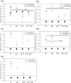
Survival of WT and mutant Y. pestis strains at 1 and 48 h p.i. in peripheral organs of mice. Groups of five animals were infected with a 250 LD50 dose of WT and mutant Y. pestis strains. At 1 and 48 h p.i., mice were euthanized, blood was drawn and organs were harvested, homogenized, serially diluted and cultured on SBA plates. Bacterial counts per gram of lung (a), liver (b), spleen (c) and heart (d) and per millilitre of blood (e) were determined for 1 h p.i. (•) and 48 h p.i. (○). Solid bars indicate the average value at 1 h p.i., while hatched bars indicate the average value at 48 h p.i. The dashed line parallel to the x axis is the limit of bacterial detection. Significant differences (Student's t test) between groups are noted.
Fig. 4.
Fig. 4.
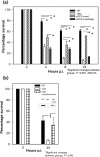
Survival of WT and mutant Y. pestis strains in vitro. Murine RAW 264.7 macrophages were infected at an m.o.i. of 1 with WT and mutant Y. pestis strains for 30 min. Monolayers were treated with 100 μg gentamicin ml−1 for 1 h, and at the indicated time points (post gentamicin), macrophages were lysed. Lysates were serially diluted and cultured on SBA plates. Colonies were counted and data were normalized to the 0 h time point. Significant differences (ANOVA, P<0.001) between groups are noted. (b) Comparison of WT- and Δlpp mutant-infected macrophages with the lpp-complemented strain of Y. pestis CO92. Significant differences (Student's t test, P<0.05) between groups are noted. The inset in (b) shows Western blot analysis data showing the presence or absence of Lpp (6.3 kDa) using specific antibodies in various strains.

References

    1. Agar, S. L., Sha, J., Foltz, S. M., Erova, T. E., Walberg, K. G., Baze, W. B., Suarez, G., Peterson, J. W. & Chopra, A. K. (2008a). Characterization of the rat pneumonic plague model: infection kinetics following aerosolization of Yersinia pestis CO92. Microbes Infect 11, 205–214. - PubMed
    1. Agar, S. L., Sha, J., Foltz, S. M., Erova, T. E., Walberg, K. G., Parham, T. E., Baze, W. B., Suarez, G., Peterson, J. W. & Chopra, A. K. (2008b). Characterization of a mouse model of plague after aerosolization of Yersinia pestis CO92. Microbiology 154, 1939–1948. - PubMed
    1. Anisimov, A. P., Bakhteeva, I. V., Panfertsev, E. A., Svetoch, T. E., Kravchenko, T. B., Platonov, M. E., Titareva, G. M., Kombarova, T. I., Ivanov, S. A. & other authors (2009). The subcutaneous inoculation of pH 6 antigen mutants of Yersinia pestis does not affect virulence and immune response in mice. J Med Microbiol 58, 26–36. - PubMed
    1. Beesley, E. D., Brubaker, R. R., Janssen, W. A. & Surgalla, M. J. (1967). Pesticins III. Expression of coagulase and mechanism of fibrinolysis. J Bacteriol 94, 19–26. - PMC - PubMed
    1. Brice, G. T., Graber, N. L., Hoffman, S. L. & Doolan, D. L. (2001). Expression of the chemokine MIG is a sensitive and predictive marker for antigen-specific, genetically restricted IFN-γ production and IFN-γ-secreting cells. J Immunol Methods 257, 55–69. - PubMed

Publication types

MeSH terms