. 2009 Oct;23(10):1668-80.
doi: 10.1210/me.2009-0066. Epub 2009 Jul 9.

The p62 P392L mutation linked to Paget's disease induces activation of human osteoclasts

Affiliations

The p62 P392L mutation linked to Paget's disease induces activation of human osteoclasts

Estelle Chamoux et al. Mol Endocrinol. 2009 Oct.

Abstract

Mutations of the gene encoding p62/SQSTM1 have been described in Paget's disease of bone (PDB), identifying p62 as an important player in osteoclast signaling. We investigated the phenotype of osteoclasts differentiated from peripheral blood monocytes obtained from healthy donors or PDB patients, all genotyped for the presence of a mutation in the p62 ubiquitin-associated domain. The cohort included PDB patients carrying or not the p62 P392L mutation and healthy donors carrying or not this mutation. Osteoclasts from PDB patients were more numerous, contained more nuclei, were more resistant to apoptosis, and had a greater ability to resorb bone than their normal counterparts, regardless of whether the p62 mutation was present or not. A strong increase in p62 expression was observed in PDB osteoclasts. The presence of the p62(P392L) gene in cells from healthy carriers conferred a unique, intermediate osteoclast phenotype. In addition, we report that two survival-promoting kinases, protein kinase Czeta and phosphoinositide-dependent protein kinase 1, were associated with p62 in response to receptor activator of NF-kappaB ligand (RANKL) stimulation in controls and before RANKL was added in PDB osteoclasts. In transfected osteoclasts derived from cord blood monocytes, the p62 P392L mutation contributed to increased activation of kinases protein kinase Czeta/lambda and phosphoinositide-dependent protein kinase 1, along with basal activation of NF-kappaB, independently of RANKL stimulation. These findings clearly indicate that the overexpression of p62 in PDB patients induces important shifts in the pathways activated by RANKL and up-regulates osteoclast functions. Moreover, the most-commonly reported p62 mutation, P392L, certainly contributes to the overactive state of osteoclasts in PDB.


Figures
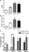
Fig. 1.
Fig. 1.
Osteoclast formation in PBMC cultures. Human PBMCs were differentiated for 21 d with RANKL and M-CSF. At the end of the culture period, cells were stained with Evan’s blue and DAPI. A, The percentage of MNCs per total cell number was evaluated. Cells containing three or more nuclei were scored as MNCs. B, The number of nuclei per MNC was counted in 20 MNCs per well. C, Bone resorption was assessed at the end of the differentiation period by toluidine blue staining of the bone slices. The resorbed surface area was measured using BioQuant Nova Image analysis software and reported graphically. Results of three to five independent experiments are expressed as mean ± sem; *, P < 0.05; **, P < 0.01; ***, P < 0.001 vs. HDwt; #, P < 0.05; ##, P < 0.01; ###, P < 0.001 vs. HDP392L; °, P < 0.05; °°°, P < 0.001 RANKL 25 ng/ml vs. 100 ng/ml. HDwt: healthy donors − no mutation in p62 gene (n = 3); HDP392L: healthy donors − p62P392L mutation (n = 3); PDBwt: PDB − no p62 mutation (n = 4); PDBP392L: PDB − with p62P392L mutation (n = 5). OC, Osteoclast.
Fig. 2.
Fig. 2.
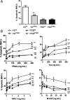
Induction of apoptosis in pagetic osteoclasts. At the end of the PBMC cultures, 24 h after the M-CSF and RANKL had been removed, apoptosis was detected by DAPI staining (A), or after adding various concentrations of hrTRAIL (10–400 ng/ml), a Fas-activating antibody (50–800 ng/ml); hrTGFβ (0.1–4 ng/ml) or M-CSF (1–50 ng/ml) where appropriate 24 h before apoptosis was assessed (B). Results of three independent experiments are expressed as the percentage of apoptotic MNCs, mean ± sem. The significance was indicated as follows: *, P < 0.05; **, P < 0.01; ***, P < 0.001 vs. untreated controls; significance of the dose-response trend-line: #, P < 0.05; ##, P < 0.01; ###, P < 0.001 vs. HDwt; ¤, P < 0.05; ¤¤, P < 0.01 HDP392L vs. PDBP392L/wt; HDwt: healthy donors − no mutation in p62 gene (n = 3); HDP392L: healthy donors − p62P392L mutation (n = 3), PDBwt: PDB − no p62 mutation (n = 5); PDBP392L: PDB − p62P392L mutation (n = 6).
Fig. 3.
Fig. 3.
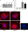
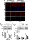
p62 protein expression in pagetic osteoclasts. Human PBMCs were differentiated for 21 d with RANKL and M-CSF. A, Whole-cell lysates of mature osteoclast cultures were subjected to SDS-PAGE followed by a Western blot with an antibody directed against p62. A control blot was done on the same membrane with an antibody directed against β-actin. B, ODs were measured with ImageJ software, and the ratio of p62 vs. actin was computed. Analyses are reported as mean ratio ± sem. *, P < 0.05 vs. HDwt. The data shown are representative of three independent experiments conducted with different combinations of samples from various donors. C, Immunofluorescence studies were performed with an antibody directed against p62 and a secondary Alexa-647 antibody (red). After washing, cells were counterstained with DAPI and mounted. Arrows, show the membrane expression of protein p62. Images presented here were representative of cells seen in five independent experiments. HDwt: healthy donors − no mutation in p62 gene (n = 3); HDP392L: healthy donors − p62P392L mutation (n = 3); PDBwt: PDB − no p62 mutation (n = 5); PDBP392L: PDB − with p62P392L mutation (n = 5). NS, Nonspecific staining.
Fig. 4.
Fig. 4.
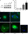
p62 protein expression in transfected osteoclasts. CBMs were differentiated for 3 wk in the presence of RANKL and M-CSF. Where appropriate, the cells were transfected on d 17 of culture with a pEGFP-C2 plasmid containing p62wt, p62P392L, or an empty pEGFP vector (EV), or were not transfected (NT). A, Cells were lysed 48 h after transfection, and whole-cell lysates were subjected to SDS-PAGE followed by a Western blot with an antibody directed against p62. A control blot was done on the same membrane with an antibody directed against β-actin. B, ODs were measured with ImageJ software, and the ratio of p62 vs. actin was computed. Analyses are reported as mean ratio ± sem. **, P < 0.01; ***, P < 0.001 vs. NT. The data shown are representative of four independent experiments. C, GFP-fused p62 was detected in transfected cells in green. As a control, endogenous levels of p62 were assessed in nontransfected osteoclasts using an anti-p62 antibody and a secondary antigoat Alex-546 antibody. After washing, the cells were counterstained with DAPI and mounted. Arrows, p62 protein was detected in the cytoplasm and membrane. The images presented here were representative of cells seen in six independent experiments. NS, Nonspecific staining.
Fig. 5.
Fig. 5.
Osteoclast phenotype in p62 transfected cells. CBMs were differentiated for 3 wk in the presence of RANKL and M-CSF. Where appropriate, the cells were transfected on d 17 of culture with a pEGFP-C2 plasmid containing p62wt, p62P392L, or an empty pEGFP vector (EV), or were not transfected (NT). A, At the end of the culture period, the number of multinucleated cells within the transfected population was evaluated. Among GFP-expressing cells, the cells containing more than three nuclei were scored as MNCs. B, The number of nuclei per MNC was counted. C, Bone resorption was assessed by toluidine blue staining. The resorbed surface area was measured using Simple PCI software, corrected for the number of MNCs under the same conditions and is reported graphically. Results are expressed as the percentage of the resorbed area. D, In apoptosis experiments, cells were deprived of RANKL and M-CSF for 24 h before apoptosis was evaluated. Apoptosis was detected by DAPI staining, which made it possible to discriminate between multinucleated transfected cells and other cells. Results are expressed as the percentage of apoptotic MNCs (or apoptotic tagged-MNCs where appropriate) over total MNCs. Results of four to six independent experiments are expressed as mean ± sem (*, P < 0.05; **, P < 0.01; ***, P < 0.001 vs. EV; #, P < 0.05 p62P392L vs. p62wt; °, P < 0.05; °°, P < 0.01 RANKL 25 vs. 100 ng/ml). OC, Osteoclast.
Fig. 6.
Fig. 6.
RANKL-induced PKCζ and p62 interactions in pagetic cells. Human PBMCs were differentiated for 21 d with RANKL and M-CSF. At the end of the culture, the cells were stimulated with RANKL (100 ng/ml) for 0, 30, 60, and 90 min. A, For HDwt and PDBwt, immunoprecipitations (IPs) were conducted with an anti-p62 antibody in whole-cell lysates. Because anti-p62 antibodies, directed against the UBA domain, were not able to efficiently precipitate the mutated p62, IPs were conducted with anti-PKCζ (panel B) or anti-P-PDK1 (panel C), for HDP392L and PDBP392L. Western blots (WB) using anti-PKCζ, anti-P-PDK1, and anti-p62 antibodies are shown. ODs were measured with ImageJ software, and the ratio of PKCζ or P-PDK1 to p62 was computed. Analyses are reported as mean ratio ± sem. ***, P < 0.001 vs. nontreated; ###, P < 0.001 vs. HDwt. The data are representative of three independent experiments. HDwt: healthy donors − no mutation in p62 gene, HDP392L: healthy donors − p62P392L mutation, PDBwt: PDB − no p62 mutation, PDBP392L: PDB − with p62P392L mutation.
Fig. 7.
Fig. 7.
RANKL-induced p62 interactions in transfected osteoclasts. At the end of CBM cultures, cells were transfected on d 17 of culture with a pEGFP-C2 plasmid containing p62wt, p62P392L, or an empty pEGFP vector (EV), or were not transfected (NT). At the end of CBM cultures, cells were stimulated with RANKL (100 ng/ml) for 0, 10, and 30 min. A, Immunoprecipitations (IPs) were conducted in nontransfected cells with an anti-p62 antibody in cell lysates. Western blots (WBs) with anti-P-PKCζ/λ, anti-P-PDK1 and anti-p62 antibodies are shown. Whole cell lysates, not submitted to IP, were run in addition to IP fractions and blotted with the same antibodies. B, In transfected cells, IPs were conducted with an anti-GFP antibody in cell lysates, to precipitate the GFP-fused p62 variant, and Western blots (WBs) with anti-P-PKCζ/λ, anti-P-PDK1, and anti-GFP antibodies are shown. C, Before immunoprecipitation, a sample of whole-cell lysate was kept and run through SDS-PAGE in parallel to the IP fractions. After transfer, PVDF membranes were blotted successively with an anti-P-PKCζ, an anti-P-PDK1, or an anti-actin antibody. The data shown are representative of four independent experiments. ODs for bands corresponding to P-PKCζ/λ and P-PDK1 were corrected with the OD obtained for bands corresponding to p62, GFP, or actin, and computed in graphical representations. Analyses are reported as mean ratio ± sem. *, P ≤ 0.05; **, P ≤ 0.01; *** , P ≤ 0.001 vs. nonstimulated cells; #, P < 0.05; ##, P < 0.01 vs. p62wt.
Fig. 8.
Fig. 8.
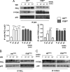
Activation of NF-κB in transfected osteoclasts. At the end of CBM cultures, cells were transfected on d 17 of culture with a pEGFP-C2 plasmid containing p62wt, p62P392L, or an empty pEGFP vector (EV), or were not transfected (NT). A, Nuclear translocation of the p50 subunit. Cells were either left untreated or stimulated with RANKL (100 ng/ml) for 30 min and then immunostained for NF-κB p50 subunit. DAPI was added at the end of the procedure to make it possible to distinguish multinucleated cells from other cells. B, Degradation of IκB. Nontransfected cells were stimulated for 5, 10, 15, 30, or 60 min with 100 ng/ml RANKL or left untreated. C, Degradation of IκB. Cells were transfected as above, then stimulated for 10 or 45 min with RANKL or left untreated. Western blots were performed with anti-IκB and antiactin antibodies. Graphic representations of average ODs measured from blots are presented. ODs for bands corresponding to IκB were corrected using the OD obtained for bands corresponding to actin. Analyses are reported as mean ratio ± sem. *, P ≤ 0.05; **, P ≤ 0.01 vs. nontreated cells. Results are representative of three experiments. WB, Western blot.

References

    1. Roodman GD, Windle JJ2005. Paget disease of bone. J Clin Invest 115:200–208 - PMC - PubMed
    1. Seitz S, Priemel M, Zustin J, Beil FT, Semler J, Minne H, Schinke T, Amling M2009. Paget’s disease of bone: histologic analysis of 754 patients. J Bone Miner Res 24:62–69 - PubMed
    1. Laurin N, Brown JP, Morissette J, Raymond V2002. Recurrent mutation of the gene encoding sequestosome 1 (SQSTM1/p62) in Paget disease of bone. Am J Hum Genet 70:1582–1588 - PMC - PubMed
    1. Morissette J, Laurin N, Brown JP2006. Sequestosome 1: mutation frequencies, haplotypes, and phenotypes in familial Paget’s disease of bone. J Bone Miner Res 21(Suppl 2):P38–P44 - PubMed
    1. Cavey JR, Ralston SH, Hocking LJ, Sheppard PW, Ciani B, Searle MS, Layfield R2005. Loss of ubiquitin-binding associated with Paget’s disease of bone p62 (SQSTM1) mutations. J Bone Miner Res 20:619–624 - PubMed

Publication types

MeSH terms

Grants and funding