. 2009 Oct 1;114(14):2888-99.
doi: 10.1182/blood-2009-01-199216. Epub 2009 Jul 9.

Lack of specific gamma-retroviral vector long terminal repeat promoter silencing in patients receiving genetically engineered lymphocytes and activation upon lymphocyte restimulation

Affiliations

Lack of specific gamma-retroviral vector long terminal repeat promoter silencing in patients receiving genetically engineered lymphocytes and activation upon lymphocyte restimulation

William R Burns et al. Blood. .

Abstract

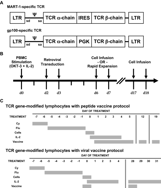
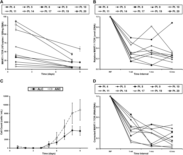
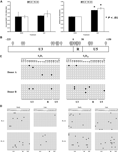
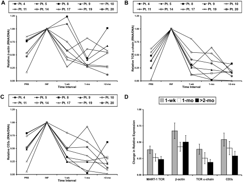
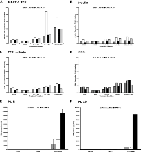
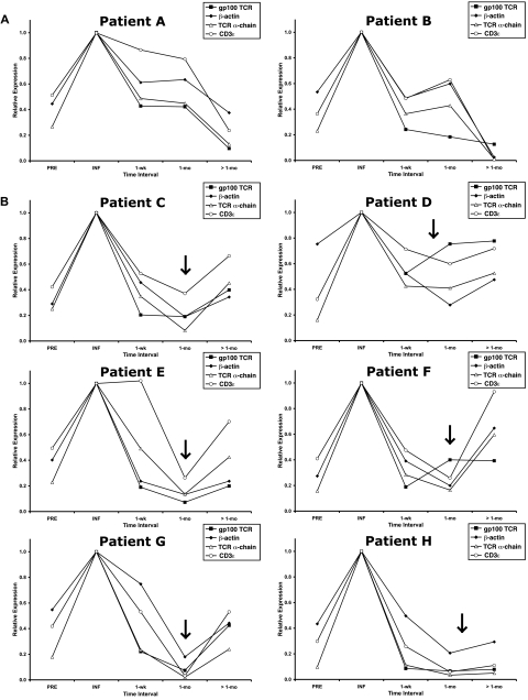
Retroviral transduction of tumor antigen-specific T-cell receptor (TCR) genes into lymphocytes redirects T cells to lyse tumors. Furthermore, adoptive transfer of these lymphocytes has mediated objective responses in patients with metastatic cancer. From 2004 to 2006, more than 40 patients were treated with autologous gene-modified lymphocytes expressing a melanoma antigen-specific TCR at the National Cancer Institute. Eighteen such patients were analyzed for persistence and gene expression in vivo. In addition, the impact of epigenetic silencing and of lymphocyte restimulation was studied. Although gene-modified lymphocytes persisted in vivo, the shutdown of TCR transgene expression was observed. Bisulfite sequencing analysis and ex vivo DNA methyltransferase inhibition demonstrated that the decrease in gene expression did not result from DNA methylation. Surprisingly, down-regulation of vector-driven transgene transcriptional activity was not vector specific but mimicked that of endogenous genes. The decrease in TCR transgene expression, however, was reversed upon lymphocyte stimulation. These data demonstrate a lack of gamma-retroviral promoter-specific gene silencing in adoptively transferred human lymphocytes and support that transgene expression is largely affected by global cellular mechanisms. The use of immunomodulatory adjuvants, eg, vaccination or cytokine therapy, for in vivo T-cell activation may help overcome this metabolic quiescence and thus augment cellular immunotherapy-based cancer therapy.


Figures

Figure 1
Figure 1
Melanoma antigen-specific TCR-encoding γ-retroviral vectors, production of autologous TCR gene-modified lymphocytes, and treatment protocols. (A) Diagram of an MSCV-based γ-retroviral vector encoding the α-chain and β-chain of a MART-1–specific TCR (top) and of an MMLV-based γ-retroviral vector encoding the α-chain and β-chain of a gp100-specific TCR (bottom). (B) Timeline depicting production of autologous TCR gene-modified lymphocytes. The infusion product was composed of cells after short-term ex vivo culture (day 6-7) with or without a portion of cells exposed to an OKT-3-based rapid expansion protocol. (C) Summary of TCR gene-modified lymphocyte protocols with administration of peptide vaccine (top) or fowlpox viral vaccine (bottom). All patients received nonmyeloablative lymphodepleting chemotherapy consisting of 2 days of cyclophosphamide (Cy) at 60 mg/kg followed by 5 days of fludarabine (Flu) at 25 mg/m2.
Figure 2
Figure 2
Levels of gene-modified cells and transgene RNA expression decrease in PBL after adoptive cell transfer in patients with metastatic melanoma. (A) DNA was isolated from cells in the treatment infusion (day 1) and subsequent peripheral blood samples of patients receiving autologous MART-1–specific TCR-transduced lymphocytes. Real-time PCR was performed to quantify the amount of transgene DNA present at each time point. Each line represents a single patient, and data points represent the mean of 3 replicates ± SD. (B) Persistence of MART-1 TCR transgene, normalized to the infusion sample (INF) at various time intervals after infusion. (C) The kinetics of immune reconstitution in the peripheral blood after the nonmyeloablative, lymphodepleting chemotherapy regimen. Adoptive cell transfer, IL-2 administration, and peptide vaccinations were initiated on day 1. Data points represent the mean of all patients in this report ± SEM. ALC indicates absolute lymphocyte count; and ANC, absolute neutrophil count. (D) MART-1–specific TCR transgene RNA expression, corrected for the level of transgene DNA, at each time interval and normalized to the infusion value (INF).
Figure 3
Figure 3
Low-level methylation of CpG residues within the LTR of proviral integrants. (A) Peripheral blood from 4 patients who received MART-1–specific TCR-transduced lymphocytes was harvested 2 months after adoptive transfer. Lymphocytes were isolated and cultured in complete media with low-dose IL-2 (60 IU/mL; MOCK) or with the addition of daily dacitabine (0.3 μmol/L; DAC). Total RNA was isolated after 3 days of ex vivo culture, reverse transcribed, and subjected to real-time PCR for the MART-1 TCR (left panel) or p21 (right panel). Each bar represents an individual patient. (B) Clustering of CpG dinucleotides within the MSCV LTR, where each oval depicts a single CpG site. Numbering is relative to the transcription start site. (C) Genomic DNA from retrovirally transduced lymphocytes of 2 donors was isolated 7 and 21 days after stimulation and bisulfite conversion was performed. A region of clustered CpG residues within the MSCV LTR were amplified by PCR and cloned. Each row represents sequencing of an individual clone, with methylated cytosine residues shown by shaded circles and nonmethylated residues by unshaded circles. (D) Bisulfite sequencing results of lymphocytes from 4 patients at early time points (1-4 weeks) and late time points (2-12 months) in vivo.
Figure 4
Figure 4
Down-regulation of transgene expression is similar to that of endogenous genes. (A) β-actin RNA expression, corrected for the level of β-actin DNA, before (PRE) and at time intervals after cell infusion were normalized to the infusion value (INF). Corrected RNA expression of the endogenous TCR α-chain (B) and CD3ϵ (C) before (PRE) and at time intervals after cell infusion were normalized to the infusion values (INF). (D) The relative, corrected MART-1–specific TCR transgene expression and the relative, corrective expression of β-actin, the endogenous TCR α-chain, and CD3ϵ at each time interval after treatment. Values are the mean of the analyzed patients ± SEM.
Figure 5
Figure 5
Up-regulation of transgenic TCR and other genes after ex vivo restimulation. Peripheral blood from 4 patients who received MART-1–specific TCR-transduced lymphocytes was harvested 2 to 10 months after adoptive transfer. Lymphocytes were isolated and cultured in complete media alone (MOCK), with exogenous IL-2 (600 IU/mL), or with IL-2 and anti-CD3/anti-CD28 beads (1 bead/cell). Total RNA was isolated at the time of in vitro culture (FRESH) or after 48 hours of ex vivo culture, reverse transcribed, and subjected to real-time PCR for (A) the MART-1–specific TCR, (B) β-actin, (C) the endogenous TCR α-chain, and (D) CD3ϵ. Each bar represents an individual patient. Peripheral blood from 2 of the 4 patients were maintained in ex vivo culture for 3 days with or without stimulation conditions and compared with the freshly thawed peripheral blood. Only those cells receiving ex vivo restimulation secreted interferon-γ and demonstrated specific recognition of MART-1 peptide-pulsed target cells (E-F).
Figure 6
Figure 6
Up-regulation of gene expression in patients after in vivo restimulation. The gp100-specific TCR transgene RNA level was corrected for the DNA level at each time interval and normalized to the infusion values (INF). RNA expression of β-actin, the endogenous TCR α-chain, and CD3ϵ before (PRE) and at time intervals after cell infusion was normalized to the infusion value (INF). (A) Patients A and B from the first cohort of this trial received IL-2 and peptide vaccination on the day of adoptive cell transfer. (B) Patients C through H from the second cohort of this trial received IL-2 with recombinant fowlpox vaccination on the day of adoptive cell transfer, as well as repeat administration of IL-2 with recombinant fowlpox vaccination approximately 1 month after adoptive cell transfer (indicated by formula image).

Comment in

  • The hidden (and lazy) TCR.
    Bonini C, Russo V. Bonini C, et al. Blood. 2009 Oct 1;114(14):2855-6. doi: 10.1182/blood-2009-07-234153. Blood. 2009. PMID: 19797529 No abstract available.

References

    1. Rosenberg SA, Restifo NP, Yang JC, Morgan RA, Dudley ME. Adoptive cell transfer: a clinical path to effective cancer immunotherapy. Nat Rev Cancer. 2008;8:299–308. - PMC - PubMed
    1. Dudley ME, Wunderlich JR, Robbins PF, et al. Cancer regression and autoimmunity in patients after clonal repopulation with antitumor lymphocytes. Science. 2002;298:850–854. - PMC - PubMed
    1. Dudley ME, Wunderlich JR, Yang JC, et al. Adoptive cell transfer therapy following non-myeloablative but lymphodepleting chemotherapy for the treatment of patients with refractory metastatic melanoma. J Clin Oncol. 2005;23:2346–2357. - PMC - PubMed
    1. Schumacher TN. T-cell-receptor gene therapy. Nat Rev Immunol. 2002;2:512–519. - PubMed
    1. Sadelain M, Rivière I, Brentjens R. Targeting tumours with genetically enhanced T lymphocytes. Nat Rev Cancer. 2003;3:35–45. - PubMed

Publication types

MeSH terms

Substances