. 2009 Sep;297(3):G461-70.
doi: 10.1152/ajpgi.90446.2008. Epub 2009 Jul 9.

Regeneration of intestinal stem/progenitor cells following doxorubicin treatment of mice

Affiliations

Regeneration of intestinal stem/progenitor cells following doxorubicin treatment of mice

Christopher M Dekaney et al. Am J Physiol Gastrointest Liver Physiol. 2009 Sep.

Abstract

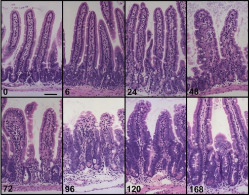
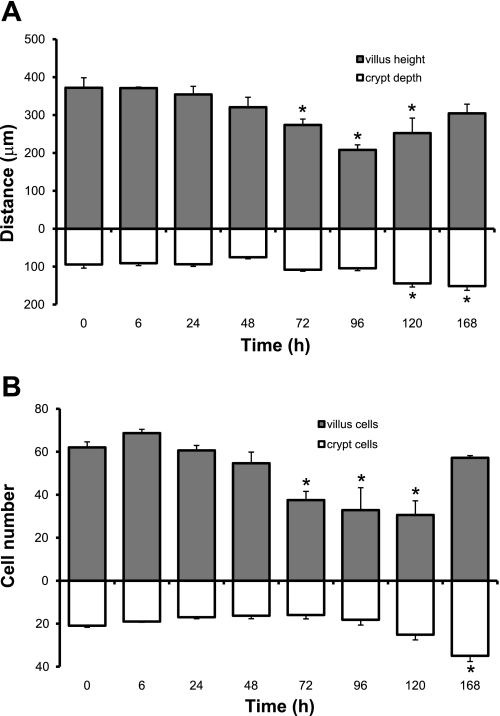
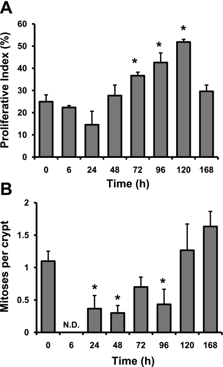
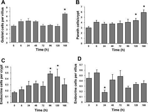
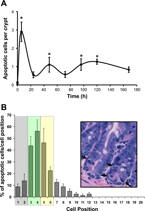
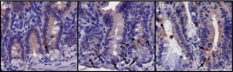
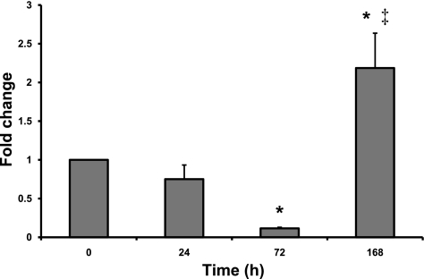
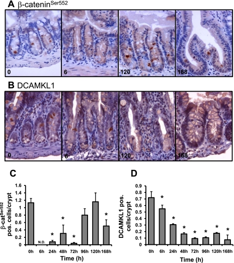
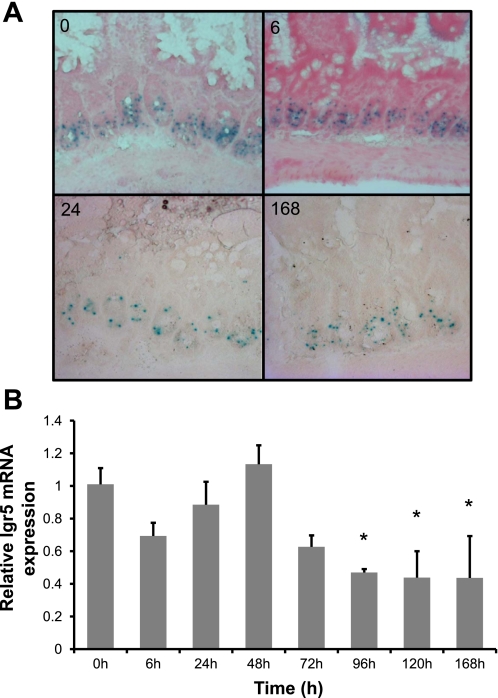
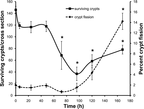
The intestinal epithelium is in a constant state of renewal. The rapid turnover of cells is fed by a hierarchy of transit amplifying and stem/progenitor cells destined to give rise to the four differentiated epithelial lineages of the small intestine. Doxorubicin (Dox) is a commonly used chemotherapeutic agent that preferentially induces apoptosis in the intestinal stem cell zone (SCZ). We hypothesized that Dox treatment would initially decrease "+4" intestinal stem cell numbers with a subsequent expansion during mucosal repair. Temporal assessment following Dox treatment demonstrated rapid induction of apoptosis in the SCZ leading to a decrease in the number of intestinal stem/progenitor cells as determined by flow cytometry for CD45(-) SP cells, and immunohistochemistry of cells positive for putative +4 stem cell markers beta-cat(Ser552) and DCAMKL1. Between 96 and 168 h postinjection, overall proliferation in the crypts increased concomitant with increases in both absolute and relative numbers of goblet, Paneth, and enteroendocrine cells. This regeneration phase was also associated with increases of CD45(-) SP cells, beta-cat(Ser552)-positive cells, crypt fission, and crypt number. We used Lgr5-lacZ mice to assess behavior of Lgr5-positive stem cells following Dox and found no change in this cell population. Lgr5 mRNA level was also measured and showed no change immediately after Dox but decreased during the regeneration phase. Together these data suggest that, following Dox-induced injury, expansion of intestinal stem cells occurs during mucosal repair. On the basis of available markers this expansion appears to be predominantly the +4 stem cell population rather than those of the crypt base.


Figures

Fig. 1.
Fig. 1.
Doxorubicin (Dox) treatment induces changes in intestinal mucosa morphology. Hematoxylin and eosin (H&E)-stained sections were evaluated for changes in the morphology of the intestinal mucosa at various times following Dox treatment. Grossly, very little damage is observed by 6 h postinjection. By 72 h, there is noticeable crypt degeneration. By 96 h villus blunting and loss of crypts becomes obvious. By 120 h crypts are clearly regenerating and villi are beginning to lengthen. Scale bar equals 50 μm.
Fig. 2.
Fig. 2.
Dox treatment alters crypt and villus morphometry. Measurements were made at 0, 6, 24, 48, 72, 96, 120, and 168 h after Dox treatment. A: villus height and crypt depth. B: cells per villus and cells per crypt. *Statistical significance compared with controls (P < 0.05) (n = 6).
Fig. 3.
Fig. 3.
Dox treatment influences proliferation. A: the number of 5′-bromo-2′-deoxyuridine (BrdU)-positive cells was scored at 0, 6, 24, 48, 72, 96, 120, and 168 h after Dox treatment and expressed as a percentage of total number of cells within a given crypt. B: the number of mitoses per crypt was scored at 0, 6, 24, 48, 72, 96, 120, and 168 h after Dox treatment by using H&E-stained sections. *Statistical significance compared with controls (P < 0.05) (n = 3). N.D., data for that time point were not detectible.
Fig. 4.
Fig. 4.
Dox treatment alters lineage allocation. Total numbers of goblet, Paneth, and enteroendocrine cells were quantified at 0, 6, 24, 48, 72, 96, 120, and 168 h after Dox treatment. A: goblet cells per half villus. B: Paneth cells per crypt. C: enteroendocrine cells per crypt. D: enteroendocrine cells per villus. *Statistical significance compared with controls (P < 0.05) (n = 5–6 for goblet and Paneth cell data; n = 3 for enteroendocrine cell data).
Fig. 5.
Fig. 5.
Dox treatment rapidly induces apoptosis centering on the putative stem cell zone. A: apoptotic cells were scored via H&E staining of jejunal sections collected at 0, 6, 24, 48, 72, 96, 120, and 168 h after Dox treatment (n = 5–6) and expressed as the number of apoptotic cells per crypt. *Statistical significance compared with zero-time controls (P < 0.05). B: the location of apoptotic bodies within particular cell positions along the crypt was recorded at the 6-h time point. Data are presented as the percent of instances a particular cell position contained an apoptotic cell. The stem/progenitor cell zone is indicated by the shaded boxes. Using the nomenclature of Scoville et al. (37), the gray box indicates the area in which crypt base columnar cells (CBCs) reside and the yellow box indicates the area containing the +4 cells. Note that the green box represents potential overlap of these 2 areas. Inset: H&E micrograph of representative crypts containing apoptotic cells (arrows).
Fig. 6.
Fig. 6.
Dox treatment induces cleavage of caspase-3. Immunohistochemistry for cleaved caspase-3 corroborated our data in Fig. 1A showing increased staining by 6 h after Dox treatment and a return to near baseline by 168 h, compared with 0-h controls. Arrows point to cleaved caspase-3-positive cells.
Fig. 7.
Fig. 7.
Dox-induced damage results in changes in CD45(−) side population (SP). Flow cytometry was used to evaluate changes in the percentage of CD45(−) SP cells 24, 72, and 168 h following Dox treatment. Data were normalized to zero-time control animals and are presented as means ± SE (n = 5). *Statistical significance compared with zero-time control (P < 0.05). ‡Statistical significance compared with all other time points (P < 0.05).
Fig. 8.
Fig. 8.
Dox treatment induces changes in the presence of β-cateninSer552 and DCAMKL1 expressing cells. Immunohistochemistry was used to identify β-cateninSer552 or DCAMKL1-positive cells 0, 6, 24, 48, 72, 96, 120, and 168 h after Dox treatment (n = 3). A: β-cateninSer552 staining at 0, 6, 120, and 168 h revealed changes in the number of positive cells that are expressed in graphic form in C. DCAMKL1 staining (B) at 0, 6, 120, and 168 h revealed changes in the number of positive cells as well, which are quantified in D. *Statistical significance compared with zero-time control (P < 0.05).
Fig. 9.
Fig. 9.
Dox treatment has no effect on LacZ activity but induces changes in mRNA expression of the putative intestinal stem cell marker Lgr5. A: staining for LacZ activity using Lgr5-lacZ mice at 0, 6, 24, and 168 h after Dox treatment demonstrates that there is little change in LacZ activity following damage and during epithelial repair. B: using real-time quantitative RT-PCR we evaluated the changes of gene expression for Lgr5 at 0, 6, 24, 48, 72, 96, 120, and 168 h after Dox treatment (n = 3). *Statistical significance compared with zero-time control (P < 0.05).
Fig. 10.
Fig. 10.
Intestinal crypt regeneration occurs following treatment with Dox. The total number of surviving intestinal crypts per cross section (solid line, primary ordinate) and the percentage of crypt fission (dashed line, secondary ordinate) were calculated in zero-time control and 6, 24, 48, 72, 96, 120, 168 h tissue post-Dox injection (n = 3). *Statistical significance compared with controls (P < 0.05).

References

    1. Ayabe T, Satchell DP, Wilson CL, Parks WC, Selsted ME, Ouellette AJ. Secretion of microbicidal alpha-defensins by intestinal Paneth cells in response to bacteria. Nat Immunol 1: 113–118, 2000. - PubMed
    1. Barker N, van de Wetering M, Clevers H. The intestinal stem cell. Genes Dev 22: 1856–1864, 2008. - PMC - PubMed
    1. Barker N, van Es JH, Kuipers J, Kujala P, van den Born M, Cozijnsen M, Haegebarth A, Korving J, Begthel H, Peters PJ, Clevers H. Identification of stem cells in small intestine and colon by marker gene Lgr5. Nature 449: 1003–1007, 2007. - PubMed
    1. Beck PL, Wong JF, Li Y, Swaminathan S, Xavier RJ, Devaney KL, Podolsky DK. Chemotherapy- and radiotherapy-induced intestinal damage is regulated by intestinal trefoil factor. Gastroenterology 126: 796–808, 2004. - PubMed
    1. Bjerknes M, Cheng H. The stem-cell zone of the small intestinal epithelium. I. Evidence from Paneth cells in the adult mouse. Am J Anat 160: 51–63, 1981. - PubMed

Publication types

MeSH terms