. 2009 Jul 10;325(5937):210-3.
doi: 10.1126/science.1173215.

A functional role for adult hippocampal neurogenesis in spatial pattern separation

Affiliations

A functional role for adult hippocampal neurogenesis in spatial pattern separation

C D Clelland et al. Science. .

Abstract

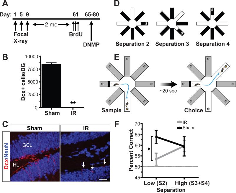
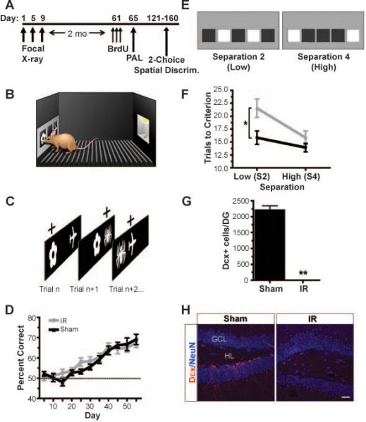
The dentate gyrus (DG) of the mammalian hippocampus is hypothesized to mediate pattern separation-the formation of distinct and orthogonal representations of mnemonic information-and also undergoes neurogenesis throughout life. How neurogenesis contributes to hippocampal function is largely unknown. Using adult mice in which hippocampal neurogenesis was ablated, we found specific impairments in spatial discrimination with two behavioral assays: (i) a spatial navigation radial arm maze task and (ii) a spatial, but non-navigable, task in the mouse touch screen. Mice with ablated neurogenesis were impaired when stimuli were presented with little spatial separation, but not when stimuli were more widely separated in space. Thus, newborn neurons may be necessary for normal pattern separation function in the DG of adult mice.


Figures

Figure 1
Figure 1. Mice with ablated neurogenesis due to focal X-irradiation show impaired spatial memory for similar, but not distinct, spatial locations in the radial arm maze
(A) Mice were irradiated 2 months prior to behavioral testing. (B,C) Irradiation significantly reduced the total numbers of immature Dcx+ cells in irradiated (IR) mice (C, right, white arrows) compared to sham controls (C, left) (independent samples t-test, t(17)=29.82 , p<0.001). (D) Pattern separation was tested using a DNMP protocol in the RAM by varying the distance between sample and correct arms: S2/low, S3 and S4/high (S=start arm). (E) Each trial consisted of a sample phase (left) and a choice phase (right). The mouse had to non-match to the novel location. (F) IR mice were impaired at low (S2) but not high (S3+S4) separations in the DNMP task. Dashed line represents chance. Error bars represent SEM. Scale bars represent 25 μm. ** p<0.01, * p<0.05. GCL indicates granule cell layer; HL, hilus.
Figure 2
Figure 2. Mice with ablated neurogenesis due to focal X-irradiation show impaired spatial discrimination for similar, but not distinct, spatial locations but not impaired associative object in-place memory in the mouse touch screen
Mice were irradiated 2 months prior to behavioral testing as in (A). Following pre-training for 7-10 days in which mice learned to nose-touch stimuli on the infrared touch screen (B) to obtain a reward, mice were trained on an associative object-in-place task (PAL) (C). For example, as in the left panel of (C), mice had to choose flower-left as a correct association over the incorrect association of plane-right in order to obtain a reward. (D) Irradiated mice (IR) learned the PAL task at the same rate as sham controls (dashed line represents chance). (E) Mice were then tested on a two-choice spatial discrimination task in which mice had to respond to the correct location (e.g., left illuminated box of left screen, E) until a criterion of 7 of 8 consecutive correct touches was recorded before reversing to the previously incorrect location (e.g., right illuminated box of left screen, E). Mice were tested on either the low separation (S2; left screen) or the high separation (S4; right screen) as depicted in (E) during each testing day. (F) IR mice exhibited significantly impaired performance at low (S2) separations but not high (S4) separations during acquisition of this task, consistent with a pattern separation also deficit observed in the first experiment (Fig. 1). (G,H) Irradiation significantly reduced the total numbers of immature Dcx+ cells in IR mice (H, right) compared to sham controls (H, left) (independent samples t-test, t(17)=18.14, p<0.001). Error bars represent SEM. Scale bars represent 50 μm. **p<0.01, * p<0.05. GCL indicates granule cell layer; HL, hilus.
Figure 3
Figure 3. Mice with decreased neurogenesis due to targeted lentiviral expression of dominant negative Wnt show impaired spatial memory for similar, but not distinct, spatial locations in the radial arm maze in a similar pattern to that seen in irradiated mice
(A,B) Dominant negative Wnt expression (B, right) significantly reduced the total numbers of immature Dcx+ cells in dnWnt mice (A) compared to GFP controls (B, left) (independent samples t-test: t(24)=3.47, p=0.002). Single channel images depicting Dcx+ cells are shown below triple channel images in (B). (C) Pattern separation was tested using a DNMP as in Fig.1. Dominant negative Wnt mice were impaired at low (S2) but not high (S3+S4) separations in the DNMP task. Error bars represent SEM. Scale bars represent 50 μm. ** p<0.01, * p<0.05. GCL indicates granule cell layer; HL, hilus.

References

    1. Leutgeb JK, Leutgeb S, Moser MB, Moser EI. Science. 2007 Feb 16;315:961. - PubMed
    1. Nakashiba T, Young JZ, McHugh TJ, Buhl DL, Tonegawa S. Science. 2008 Feb 29;319:1260. - PubMed
    1. Bakker A, Kirwan CB, Miller M, Stark CE. Science. 2008 Mar 21;319:1640. - PMC - PubMed
    1. Marr D. Philos Trans R Soc Lond B Biol Sci. 1971 Jul 1;262:23. - PubMed
    1. Jung MW, McNaughton BL. Hippocampus. 1993 Apr;3:165. - PubMed

Publication types