Early ontogeny of the olfactory organ in a basal actinopterygian fish: polypterus
- PMID: 19590178
- PMCID: PMC2746032
- DOI: 10.1159/000228162
Early ontogeny of the olfactory organ in a basal actinopterygian fish: polypterus
Abstract
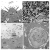
The present study employed light and electron microscopic methods to investigate the ontogenetic origin of the olfactory organ in bichirs (Cladistia: Polypteridae) and explore its evolution among osteichthyans. In former studies we demonstrated that in teleosts a subepidermal layer gives rise to the olfactory placode which in turn builds all types of olfactory cells (basal, receptor, supporting, ciliated non-sensory cells). In contrast, the olfactory placodes in sturgeons (Chondrostei: Acipenseridae) as well as in the clawed frog Xenopus laevis (Anura: Pipidae) originate from two different layers. Receptor neurons derive from cells of the subepidermal (sensory) layer and supporting cells from epidermal cells. As sturgeons and amphibians in some characters show a more primitive condition than teleosts, we extended our study to Polypterus to allow for an approach at the basic osteichthyan pattern. In Polypterus, an internal lumen occurs in early ontogenetic stages surrounded by the epithelium of the olfactory placode. Two different populations of supporting cells follow one another: a primary population derives from the subepidermal layer. Later supporting cells develop from epidermal cells by transdifferentiation. The primary opening of the internal lumen to the exterior develops by invagination from the epidermal surface and simultaneously by a counter-directed process of cell dissociation and fragmentation inside the olfactory placode. Our results indicate the following features to be plesiomorphic actinopterygian character states: The primary olfactory pit (prospective olfactory cavity) is formed by invagination of the epidermal and the subepidermal layer (as in Acipenser and Xenopus). The incurrent and excurrent nostrils derive from a single primary opening which elongates and is then separated by an epidermal bridge into the two external openings (as in Acipenser and many teleosts). The olfactory epithelium derives from an epidermal and a subepidermal layer (as in Acipenser and Xenopus). Apomorphic (derived actinopterygian) features are: (1) an internal lumen as primordium of the future olfactory chamber; (2) a subepidermal layer gives rise to the olfactory epithelium and its constituents (Polypterus and teleosts). As to the origin of the olfactory supporting cells in Polypterus we assume a combination of plesiomorphic and apomorphic characters. We conclude that Acipenser and Xenopus exhibit the most widely distributed features among basal osteognathostomes and thus ancestral character states in the development of the olfactory organs.
Copyright 2009 S. Karger AG, Basel.
Figures


Similar articles
-
Early development of the olfactory organ in sturgeons of the genus Acipenser: a comparative and electron microscopic study.Anat Embryol (Berl). 2003 Apr;206(5):357-72. doi: 10.1007/s00429-003-0309-6. Epub 2003 Mar 21. Anat Embryol (Berl). 2003. PMID: 12684762
-
Development of the olfactory organ in the zebrafish, Brachydanio rerio.J Comp Neurol. 1993 Jul 8;333(2):289-300. doi: 10.1002/cne.903330213. J Comp Neurol. 1993. PMID: 8345108
-
Ultrastructural and developmental anatomy of the peripheral olfactory organs of Dicentrarchus labrax inhabiting Egyptian Mediterranean water.Open Vet J. 2025 Feb;15(2):939-953. doi: 10.5455/OVJ.2025.v15.i2.43. Epub 2025 Feb 28. Open Vet J. 2025. PMID: 40201804 Free PMC article.
-
Neural crest and placode contributions to olfactory development.Curr Top Dev Biol. 2015;111:351-74. doi: 10.1016/bs.ctdb.2014.11.010. Epub 2015 Jan 20. Curr Top Dev Biol. 2015. PMID: 25662265 Review.
-
The cranial nerves of the Senegal bichir, Polypterus senegalus [osteichthyes: actinopterygii: cladistia].Brain Behav Evol. 1996;47(2):55-102. doi: 10.1159/000113229. Brain Behav Evol. 1996. PMID: 8866706 Review.
Cited by
-
Olfactory Sensory Neuron Morphotypes in the Featherback Fish, Notopterus notopterus (Osteoglossiformes: Notopteridae).Ann Neurosci. 2014 Apr;21(2):51-6. doi: 10.5214/ans.0972.7531.210205. Ann Neurosci. 2014. PMID: 25206061 Free PMC article.
References
-
- Bartsch P, Britz R. A single micropyle in the eggs of the most primitive living actinopterygian fish Polypterus. J Zool Lond. 1997;241:589–592.
-
- Bartsch P, Gemballa S, Piotrowski T. The embryonic and larval development of Polypterus senegalus Cuvier, 1829: its staging with reference to external and skeletal features, behaviour and locomotory habits. Acta Zool Stockholm. 1997;78:309–328.
-
- Bemis WE, Grande L. Early development of the actinopterygian head. I. External development and staging of the paddlefish Polyodon spathula. J Morphol. 1992;213:47–83. - PubMed
-
- Berliner K. Die Entwicklung des Geruchsorgans der Selachier. Arch mikrosk Anat EntwMech. 1902;60:386–406.
-
- Bertmar G. On the ontogeny and homology of the choanal tubes and choanae in Urodela. Acta Zool Stockholm. 1966;47:43–59.
Publication types
MeSH terms
Grants and funding
LinkOut - more resources
Full Text Sources
Research Materials

