. 2009;113(1):e26-34.
doi: 10.1159/000228080. Epub 2009 Jul 9.

Aldosterone induces apoptosis in rat podocytes: role of PI3-K/Akt and p38MAPK signaling pathways

Affiliations

Aldosterone induces apoptosis in rat podocytes: role of PI3-K/Akt and p38MAPK signaling pathways

Cheng Chen et al. Nephron Exp Nephrol. 2009.

Abstract

Background: Podocytes play a critical role in the pathogenesis of glomerulosclerosis. Increasing evidence suggests that aldosterone (ALD) is involved in the initiation and progression of glomerular damage. It is, however, unknown whether there is a direct injurious effect of ALD on podocytes. Therefore, in the present study, we evaluated the effect of ALD on podocyte apoptosis and studied the role of phosphatidylinositol 3-kinase/Akt (PI3-K/Akt) and p38 mitogen-activated protein kinase (p38MAPK) signaling pathways in this process.

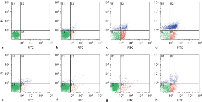
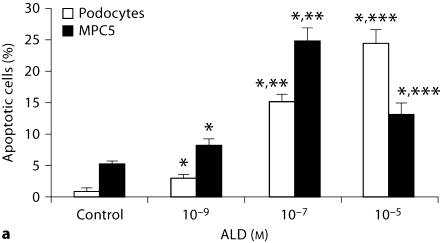
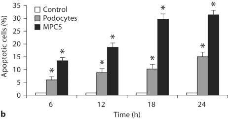
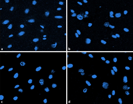
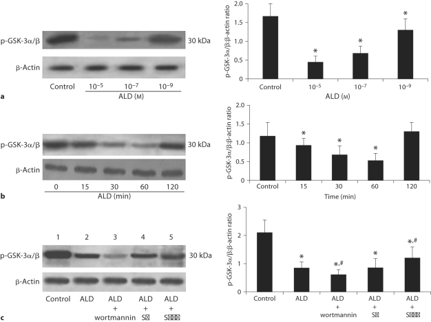
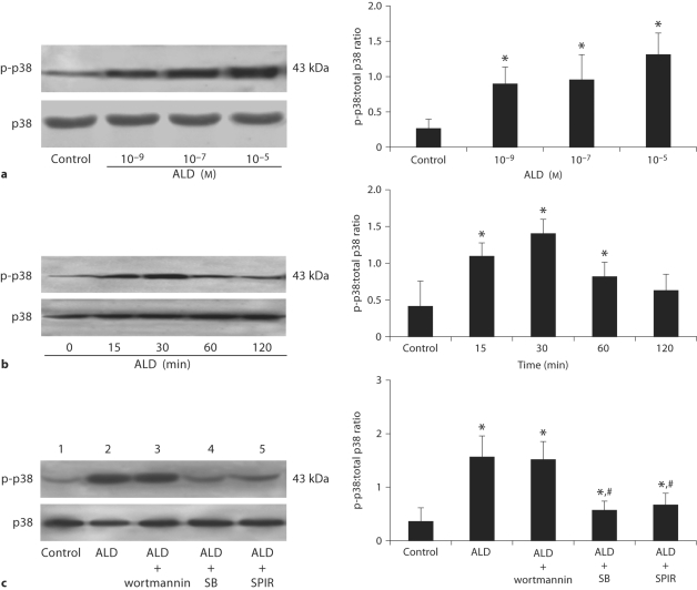
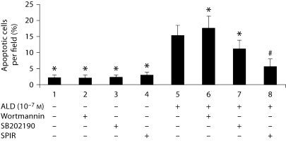
Methods: Podocytes were incubated in media containing either buffer or increasing concentrations of ALD (10(-9) approximately 10(-5)M) for variable time periods. The cells were also treated with either wortmannin (inhibitor of PI3-K, 100 nM), SB202190 (SB20, inhibitor of p38MAPK, 10 microM) or buffer. All treatments were performed with or without ALD (10(-7)M) for 24 h. At the end of the incubation period, apoptosis was evaluated by cell nucleus staining and flow cytometric analyses. Activation of PI3-K/Akt and p38MAPK phosphorylation of cultured rat podocytes was evaluated by performing Akt kinase assay and Western blot, respectively.

Results: Apoptosis of cultured rat podocytes was induced by ALD in a dose- and time-dependent manner. ALD inhibited the activity of PI3-K/Akt and increased the activation of p38MAPK. PI3-K/Akt activity was further inhibited by the addition of wortmannin to the cells in the presence of ALD. This was accompanied by a significant increase in apoptosis. ALD-induced p38MAPK phosphorylation and apoptosis were inhibited when the cells were pretreated with SB20. Furthermore, treatment with spironolactone not only attenuated the proapoptotic effect of ALD, but also significantly reversed its effects on PI3-K/Akt and p38MAPK signaling pathways.

Conclusion: ALD induces apoptosis in rat podocytes through inhibition of PI3-K/Akt and stimulation of p38 MAPK signaling pathways. Spironolactone attenuates ALD-induced podocyte apoptosis, thereby positioning this compound as a potential promising target of intervention in human renal damage.


Figures

Fig. 1.
Fig. 1.
Dose- and time-dependent apoptosis induced by ALD in podocytes. a–d Dose-response effect of ALD on podocyte apoptosis. Equal numbers of subconfluent podocytes were incubated in media containing either buffer (as a control; a) or variable concentrations of ALD (10−9 to 10−5M, b–d) for 24 h. At the end of the incubation period, the extent of apoptosis was determined by flow cytometry using PI for hypodiploid DNA and annexin V double staining. X-axis represents FITC staining and y-axis represents PI staining. Left lower quadrant means annexin V-FITC and PI negative. Right lower quadrant means annexin-V FITC positive and PI negative. Right superior quadrant means annexin V-FITC and PI positive. Left superior quadrant means PI positive. Viable cells were annexin V-FITC and PI negative, cells in early-stage apoptosis were annexin-V FITC positive and PI negative, cells in late-stage apoptosis and already dead were annexin V-FITC and PI positive. e–h Time response effects of ALD on podocyte apoptosis. Equal numbers of subconfluent podocytes were treated with ALD (10−7M) for 6 (e), 12 (f), 18 (g) and 24 h (h). At the confining point, cells were stained as described above.
Fig. 2.
Fig. 2.
Dose-response (a) and time course (b) effect of ALD on primary cultured podocyte and MPC5 cell apoptosis. a Equal numbers of cells were incubated in media containing either buffer (control) or 10−9 and 10−5M ALD for 24 h. At the end of the incubation period, cells were stained for apoptosis. The concentration of 10−5M ALD was so high to MPC5 cells that it caused necrosis directly. Values are mean ± SE of 3 sets of experiments, each carried out in triplicate. ∗ p < 0.05 vs. control; ∗∗ p < 0.05 vs. 10−9M ALD; ∗∗∗ p < 0.05 vs. 10−7M ALD. b Time course effect of ALD (10−7M) on primary cultured podocyte and MPC5 cell apoptosis. Equal numbers of cells were incubated in media containing either buffer (control) or 10−7M ALD for variable time periods (6, 12, 18, and 24 h). At the end of the incubation period, cells were stained for apoptosis. ∗ p < 0.05, compared with respective controls.
Fig. 2.
Fig. 2.
Dose-response (a) and time course (b) effect of ALD on primary cultured podocyte and MPC5 cell apoptosis. a Equal numbers of cells were incubated in media containing either buffer (control) or 10−9 and 10−5M ALD for 24 h. At the end of the incubation period, cells were stained for apoptosis. The concentration of 10−5M ALD was so high to MPC5 cells that it caused necrosis directly. Values are mean ± SE of 3 sets of experiments, each carried out in triplicate. ∗ p < 0.05 vs. control; ∗∗ p < 0.05 vs. 10−9M ALD; ∗∗∗ p < 0.05 vs. 10−7M ALD. b Time course effect of ALD (10−7M) on primary cultured podocyte and MPC5 cell apoptosis. Equal numbers of cells were incubated in media containing either buffer (control) or 10−7M ALD for variable time periods (6, 12, 18, and 24 h). At the end of the incubation period, cells were stained for apoptosis. ∗ p < 0.05, compared with respective controls.
Fig. 3.
Fig. 3.
Hoechst-33324 staining of podocytes. Representative fluorescence microscopy of Hoechst-33342 staining at 24 h after incubation of podocytes in media containing either buffer (as control; a) or variable concentrations of ALD (10−9, 10−7, 10−5M; b–d, respectively). a In the control group, 1.81% of podocytes underwent apoptosis. b–d ALD increased podocyte apoptosis at most to 24.44% in ten random nonoverlapping fields. b Bright and rippled nuclei were observed, indicating the nature of early stage of apoptosis. c, d High condensation and marginalization became increasingly clear in the nuclei, indicating the nature of late stage of apoptosis.
Fig. 4.
Fig. 4.
Western blot assay for Akt in podocytes. a ALD decreases activation of p-GSK-3α/β which can reflect the activation of Akt. Podocytes were treated with either buffer (as control) or variable concentrations of ALD (10−5 to 10−9M). Cell lysates were prepared and used for measuring phosphorylated levels of GSK-3α/β,or β-actin immunoblot using respective antibodies. b Effects of ALD on Akt expression in podocytes. Podocytes were treated with or without ALD (10−7M) for the indicated times (0 as control). At each time point, whole cell lysates were prepared and used for p-GSK-3α/β or β-actin immunoblot using respective antibodies. c Effects of wortmannin, SB202190 and SPIR on ALD-induced Akt expression in podocytes. Podocytes were pretreated for 1 h with wortmannin (lane 3), SB202190 (lane 4), or SPIR (lane 5) before being treated with buffer (as control, lane 1) or ALD (10−7M, lanes 2−5) for 60 min. Whole cell lysates were prepared and analyzed by immunoblot to detect p-GSK-3α/β. ∗ p < 0.05 vs. control group; # p < 0.05 vs. ALD group.
Fig. 5.
Fig. 5.
Western blot assay for phosphorylated p38MAPK in podocytes. Podocytes were treated with either 10−9, 10−7, or 10−5M ALD (a) or 10−7M ALD (b) for 0, 15, 30, 60, 120 min. Cell lysates were prepared and immunoblotted to measure phosphorylated and total levels of p38MAPK as indicated. c Effects of wortmannin, SB202190 and SPIR on ALD-induced p38MAPK expression in podocytes. Podocytes were pretreated for 1 h with wortmannin (lane 3), SB202190 (lane 4), or SPIR (lane 5) before being treated with buffer (as control, lane 1) or ALD (10−7M, lanes 2−5) for 30 min. Whole cell lysates were prepared and analyzed by immunoblot to detect p-p38MAPK and total p38MAPK. ∗ p < 0.05 vs. control group; # p < 0.05 vs. ALD group.
Fig. 6.
Fig. 6.
Effect of wortmannin, SB202190 and SPIR on ALD-induced podocyte apoptosis. Equal numbers of cells were pretreated for 1 h with wortmannin, SB202190 and SPIR, respectively, before treatment with or without ALD (10−7M) for 24 h. At the end of the experiments, cells were stained for apoptosis. ∗ p < 0.05 vs. ALD group.

References

    1. Espiner E, Nicholls M. The Renin Angiotensin System. London: Gower Medical Publishing; 2002. pp. 1–33.
    1. Brown NJ. Aldosterone and end-organ damage. Curr Opin Nephrol Hypertens. 2005;14:235–241. - PubMed
    1. Miric G, Dallemagne C, Endre Z, et al. Reversal of cardiac and renal fibrosis by pirfenidone and spironolactone in streptozotocin-diabetic rats. Br J Pharmacol. 2001;133:687–694. - PMC - PubMed
    1. Rocha R, Chander PN, Zuckerman A, et al. Role of aldosterone in renal vascular injury in stroke-prone hypertensive rats. Hypertension. 1999;33:232–237. - PubMed
    1. Sun Y, Zhang J, Lu L, et al. Aldosterone-induced inflammation in the rat heart: role of oxidative stress. Am J Pathol. 2002;161:1773–1781. - PMC - PubMed

Publication types

MeSH terms