. 2009 Sep 10;23(14):1799-806.
doi: 10.1097/QAD.0b013e32832ec1dc.

Expression of latent human immunodeficiency type 1 is induced by novel and selective histone deacetylase inhibitors

Affiliations

Expression of latent human immunodeficiency type 1 is induced by novel and selective histone deacetylase inhibitors

Nancie M Archin et al. AIDS. .

Abstract

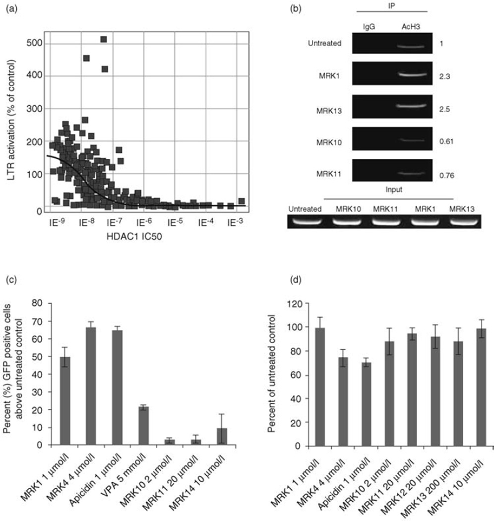
Objectives: A family of histone deacetylases (HDACs) mediates chromatin remodeling, and repression of gene expression. Deacetylation of histones within the HIV-1 long terminal repeat (LTR) by HDACs plays a key role in the maintenance of latency, whereas acetylation of histones about the LTR is linked to proviral expression and escape of HIV from latency. Global HDAC inhibition may adversely affect host gene expression, leading to cellular toxicities. Potent inhibitors selective for HDACs that maintain LTR repression could be ideal antilatency therapeutics.

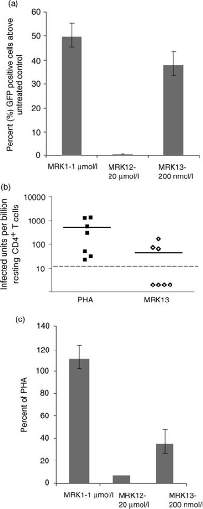
Methods: We investigated the ability of selective HDAC inhibitors to de-repress the HIV-1 LTR in both a cell line model of latency and in resting CD4 T cells isolated from patients who were aviremic on antiretroviral therapy (ART).

Results: We found that inhibition of class I HDACs increased acetylation of histones at the LTR, but that LTR chromatin was unaffected by class II HDAC inhibitors. In a latently infected cell line, inhibitors selective for class I HDACs were more efficient activators of the LTR than inhibitors that target class II HDACs. Class I HDAC inhibitors were strikingly efficient inducers of virus outgrowth from resting CD4 T cells of aviremic patients, whereas HIV was rarely recovered from patient's cells exposed to class II HDAC inhibitors.

Conclusions: Further development of selective HDAC inhibitors as part of a clinical strategy to target persistent HIV infection is warranted.


Figures

Fig. 1
Fig. 1. Class I and nonselective HDAC inhibition induces LTR expression more effectively than class II HDAC inhibitors
(a) HIV LTR expression is increased by HDAC inhibitors with increasing inhibitory potential for HDAC1. Hela P4/R5 cells were seeded and incubated for 24 h, followed by addition of HDAC inhibitors. β-galactosidase activity was measured after 24 h incubation with inhibitors. LTR activation is reported as the percentage β-galactosidase activity in treated cells over untreated control. (b) Class I but not class II HDAC inhibitors increase acetylation of Nuc1 at the HIV-1 LTR. J89 cells were treated with media, or HDAC inhibitors: MRK 1 or MRK 13 at class I HDAC1 IC50 (500 nmol/l and 300 nmol/l, respectively), MRK 10 or MRK 11 at class II HDAC4 IC50 (200 nmol/l and 500 nmol/l, respectively). Cells were treated for 4 h and assayed by chromatin immunoprecipitation with control rabbit IgG or anti-acetylated H3. DNA products of ChIP were quantitated in triplicate by real-time PCR. Assays are representative of three independent experiments, and real-time quantitation of the fold change relative to untreated control is shown. Only class I inhibitors demonstrate significant increases in histone H3 acetylation. (c) Class I and nonselective HDAC inhibition induces HIV expression. J89 cells were incubated overnight with the indicated concentrations of the HDACi. LTR-driven GFP production was measured by flow cytometry as described in methods. The data presented are the mean ± SE of three independent experiments. (d) Toxicity of HDAC inhibitors on J89 cells at concentrations used to measure promoter activation. J89 cells were cultured in the absence or presence of the indicated inhibitors for 24 h at the concentrations displayed. 3-(4,5-Dimethylthiazol-2-yl)-2,5-diphenyltetrazolium bromide (MTT) assays were performed in triplicate. The percentage of proliferating cells was calculated compared to cells cultured in standard media. ChIP, chromatin immunoprecipitation; GFP, green florescence protein; HDAC, histone deacetylase; LTR, long terminal repeat.
Fig. 2
Fig. 2. Recovery of replication-competent HIV from the resting CD4+ T cells of aviremic patients treated with HDAC inhibitors: MRK 1 (class I HDACs 1, 2, and 3 and class II HDAC6), VPA (nonselective), or MRK 10 or 11 (class II HDACs)
Patient cells were subjected to maximum mitogen stimulation or exposed to 2 µmol/l MRK 1, 40 µmol/l VPA, 2 µmol/l MRK 10 and 10 µmol/l MRK 11 for 24 h. Cells were washed and co-cultured with CD8-depleted PBMC as detailed in methods. Frequency of virus outgrowth from cells treated with HDACi was compared to the frequency of outgrowth from maximally stimulated cells (PHA). Each icon represents independent studies of patient cell samples; patient samples were simultaneously tested with mitogen or HDACi on the same day. Dashed lines indicate the limit of detection of the assay. HDAC, histone deacetylase; PBMC, peripheral blood mononuclear cell; VPA, valproic acid.
Fig. 3
Fig. 3. Recovery of replication-competent HIV from resting CD4+ T cells of aviremic patients treated with Apicidin and MRK 4, inhibitors selective only for the class I HDACs 1, 2, and 3. Patient cells were subjected to maximum mitogen stimulation or exposed to 4 µmol/l MRK 4 or 1 µmol/l MRK 6 for 24 h
Cells were washed and co-cultured with CD8-depleted PBMC as described in methods. Frequency of virus outgrowth from cells treated with HDACi was compared to outgrowth after maximal mitogen activation by PHA. Each icon represents independent studies of patient cell samples; patient samples were simultaneously tested with mitogen or HDACi on the same day. Dashed lines indicate the limit of detection of the assay. HDAC, histone deacetylase; PBMC, peripheral blood mononuclear cell.
Fig. 4
Fig. 4. HIV expression and outgrowth is induced by inhibitors targeting HDACs 1, 2, and 3, but viral outgrowth is markedly improved by the tandem inhibition of HDAC1–2 and the class II HDAC6
(a) J89 cells were incubated overnight with 1 µmol/l MRK 1 (selective for HDAC1, 2, 3 and 6), 2–20 µmol/l MRK 12 (HDAC1 and 2) or 200 nmol/l MRK 13 (HDAC1, 2, 3). LTR-driven GFP production was measured by flow cytometry as described in methods. The data presented are the mean ± SE of three independent experiments. (b) Resting CD4+ T cells from patients were subjected to maximum mitogen stimulation or exposed to 200–300 nmol/l MRK 13 for 24 h. Cells were washed and co-cultured with CD8-depleted PBMC as described in methods. Frequency of virus outgrowth from cells treated with MRK 13 was compared to outgrowth after maximal mitogen activation. Each icon represents independent studies of patient cell samples; patient samples were simultaneously tested with mitogen or HDACi on the same day. Dashed lines indicate the limit of detection of the assay. (c) Frequency of virus outgrowth from resting CD4+ T cells of aviremic HIV+ patients exposed to 2 µmol/l MRK 1, 2–20 µmol/l MRK 12 and 300 nmol/l MRK 13 are shown as a percentage of outgrowth obtained from corresponding maximal mitogen activation by PHA. MRK 1, n = 8; MRK 12, n = 2; MRK 13, n = 7. GFP, green florescence protein; HDAC, histone deacetylase; LTR, long terminal repeat; PBMC, peripheral blood mononuclear cell.

Similar articles

Cited by

References

    1. Chun TW, Stuyver L, Mizell SB, Ehler LA, Mican JA, Baseler M, et al. Presence of an inducible HIV-1 latent reservoir during highly active antiretroviral therapy. Proc Natl Acad Sci U S A. 1997;94:13193–13197. - PMC - PubMed
    1. Finzi D, Hermankova M, Pierson T, Carruth LM, Buck C, Chaisson RE, et al. Identification of a reservoir for HIV-1 in patients on highly active antiretroviral therapy. Science. 1997;278:1295–1300. - PubMed
    1. Wong JK, Hezareh M, Gunthard HF, Havlir DV, Ignacio CC, Spina CA, et al. Recovery of replication-competent HIV despite prolonged suppression of plasma viremia. Science. 1997;278:1291–1295. - PubMed
    1. Pazin MJ, Sheridan PL, Cannon K, Cao Z, Keck JG, Kadonaga JT, et al. NF-kappa B-mediated chromatin reconfiguration and transcriptional activation of the HIV-1 enhancer in vitro. Genes Dev. 1996;10:37–49. - PubMed
    1. Van Lint C, Emiliani S, Ott M, Verdin E. Transcriptional activation and chromatin remodeling of the HIV-1 promoter in response to histone acetylation. EMBO J. 1996;15:1112–1120. - PMC - PubMed

Publication types