. 2009 Aug;18(8):1674-83.
doi: 10.1002/pro.181.

Exploring functional roles of multibinding protein interfaces

Affiliations

Exploring functional roles of multibinding protein interfaces

Manoj Tyagi et al. Protein Sci. 2009 Aug.

Abstract

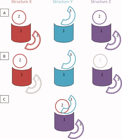
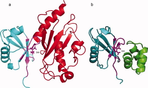
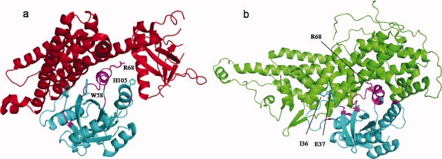
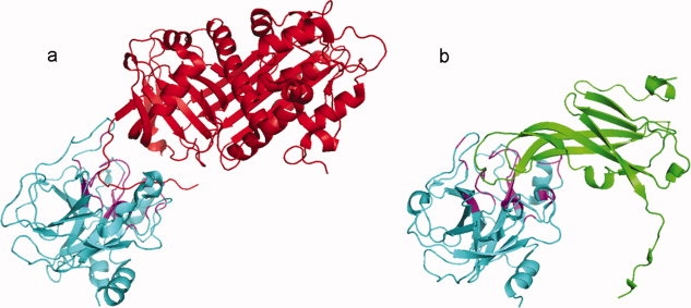
Cellular processes are highly interconnected and many proteins are shared in different pathways. Some of these shared proteins or protein families may interact with diverse partners using the same interface regions; such multibinding proteins are the subject of our study. The main goal of our study is to attempt to decipher the mechanisms of specific molecular recognition of multiple diverse partners by promiscuous protein regions. To address this, we attempt to analyze the physicochemical properties of multibinding interfaces and highlight the major mechanisms of functional switches realized through multibinding. We find that only 5% of protein families in the structure database have multibinding interfaces, and multibinding interfaces do not show any higher sequence conservation compared with the background interface sites. We highlight several important functional mechanisms utilized by multibinding families. (a) Overlap between different functional pathways can be prevented by the switches involving nearby residues of the same interfacial region. (b) Interfaces can be reused in pathways where the substrate should be passed from one protein to another sequentially. (c) The same protein family can develop different specificities toward different binding partners reusing the same interface; and finally, (d) inhibitors can attach to substrate binding sites as substrate mimicry and thereby prevent substrate binding.


Figures

Figure 1
Figure 1
Procedure of mapping of multibinding interaction interface on the domain family. At the first stage (row A), all interactions involving a given domain family “1” are collected, which in this case involve interactions with domains “2” and “3” from structures “X”, “Y,” and “Z”. The interactions are categorized into CBMs and interaction 1:3 is found to occur in two unique CBMs (the second of which is distinguished with the compound border). At the second stage, the representatives for each CBM of each interaction are chosen (row B). At the third stage, all structures X, Y, and Z are structurally superimposed and interfaces from all CBMs are mapped onto a template of domain “1” (row C). [Color figure can be viewed in the online issue, which is available at www.interscience.wiley.com.]
Figure 2
Figure 2
Interface overlap ratio between homo- and heterodimers (black bar) and between different heterodimers (gray bar).
Figure 3
Figure 3
Amino acid composition at multibinding sites.
Figure 4
Figure 4
Functional switch between different functional pathways. (a) Interaction between histidine-containing phosphocarrier protein domain (HPr, cd00367, 1RZRY, cyan) and transcription regulators (pfam00532, 1RZRD, red). (b) Interaction between Hpr domain (cd00367, 3EZAB, cyan) and PEP-utilizing enzymes (pfam05524, 3EZAA, green). An overlap of 64% is observed on the Hpr domain interface between the above two interactions, as shown in magenta color. Side chains of key interface residues like H15 and S46 are also shown. [Color figure can be viewed in the online issue, which is available at www.interscience.wiley.com.]
Figure 5
Figure 5
Multibinding interface on ubiquitin domain. (a) Interaction between ubiquitin domain, UBQ (cd01803, 1ZGUB, cyan) and ubiquitin-conjugating enzyme E2 (smart00212, 1ZGUA, red). (b) Interactions between ubiquitin domain (cd01803, 2G3QB, cyan) and ubiquitin-associated domain, UBA (pfam00627, 2G3QA, green). An overlap of 55% is observed on UBQ domain interface and is shown in magenta. Key interface residue I44 on promiscuous interface is shown with the side chain. [Color figure can be viewed in the online issue, which is available at www.interscience.wiley.com.]
Figure 6
Figure 6
Specific binding on promiscuous site. (a) Interaction between Rho GTPase domain (cd00157, 1X86F, cyan) and GEF for Rho-like GTPases (RhoGEF, cd00160, 1X86E, red). (b) Interaction between Ras GTPase domain (smart00173, 1NVVR, cyan) and GEF for Ras-like GTPases (RasGEF, cd00155, !NVVS, green). GTPase domain in both interactions shares similar interface region and overlap on 50% of the interface residues, shown in magenta color. Specific interface residues in each of the interactions are shown with the side chains, for example, I36 and E37 on Ras GTPases and W58 and H105 on Rho GTPases. Residues like R68 are shared by both interactions and are part of the critical region of the interface site. Families cd00157 and smart00173 belong to one superfamily cluster (see Methods). [Color figure can be viewed in the online issue, which is available at www.interscience.wiley.com.]
Figure 7
Figure 7
Inhibitors mimic substrate binding in serine proteases. (a) Interaction between trypsin-like serine protease (cd00190, 1TB6H, cyan) and antithrombin from serpin superfamily (cd02045, 1TB6I, red). (b) Interaction between serine protease (cd00190, 1XX9B, cyan) and protease inhibitor ecotin (cd00242, 1XX9D, green). The active site of proteases is targeted by different inhibitors; both of them mimic the substrate resulting in inactive proteases. In this case, 67% of the interface residues are shared between two inhibitors as shown in magenta color. [Color figure can be viewed in the online issue, which is available at www.interscience.wiley.com.]

References

    1. Gavin AC, Bosche M, Krause R, Grandi P, Marzioch M, Bauer A, Schultz J, Rick JM, Michon AM, Cruciat CM, Remor M, Hofert C, Schelder M, Brajenovic M, Ruffner H, Merino A, Klein K, Hudak M, Dickson D, Rudi T, Gnau V, Bauch A, Bastuck S, Huhse B, Leutwein C, Heurtier MA, Copley RR, Edelmann A, Querfurth E, Rybin V, Drewes G, Raida M, Bouwmeester T, Bork P, Seraphin B, Kuster B, Neubauer G, Superti-Furga G. Functional organization of the yeast proteome by systematic analysis of protein complexes. Nature. 2002;415:141–147. - PubMed
    1. Krogan NJ, Peng WT, Cagney G, Robinson MD, Haw R, Zhong G, Guo X, Zhang X, Canadien V, Richards DP, Beattie BK, Lalev A, Zhang W, Davierwala AP, Mnaimneh S, Starostine A, Tikuisis AP, Grigull J, Datta N, Bray JE, Hughes TR, Emili A, Greenblatt JF. High-definition macromolecular composition of yeast RNA-processing complexes. Mol Cell. 2004;13:225–239. - PubMed
    1. Krause R, von Mering C, Bork P, Dandekar T. Shared components of protein complexes—versatile building blocks or biochemical artefacts= Bioessays. 2004;26:1333–1343. - PubMed
    1. Maniatis T, Reed R. An extensive network of coupling among gene expression machines. Nature. 2002;416:499–506. - PubMed
    1. Beckett D. Functional switches in transcription regulation; molecular mimicry and plasticity in protein-protein interactions. Biochemistry. 2004;43:7983–7991. - PubMed

Publication types

LinkOut - more resources