. 2009 Sep;27(9):2362-70.
doi: 10.1002/stem.163.

Neural stem cell transplantation benefits a monogenic neurometabolic disorder during the symptomatic phase of disease

Affiliations

Neural stem cell transplantation benefits a monogenic neurometabolic disorder during the symptomatic phase of disease

Mylvaganam Jeyakumar et al. Stem Cells. 2009 Sep.

Abstract

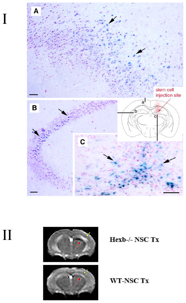
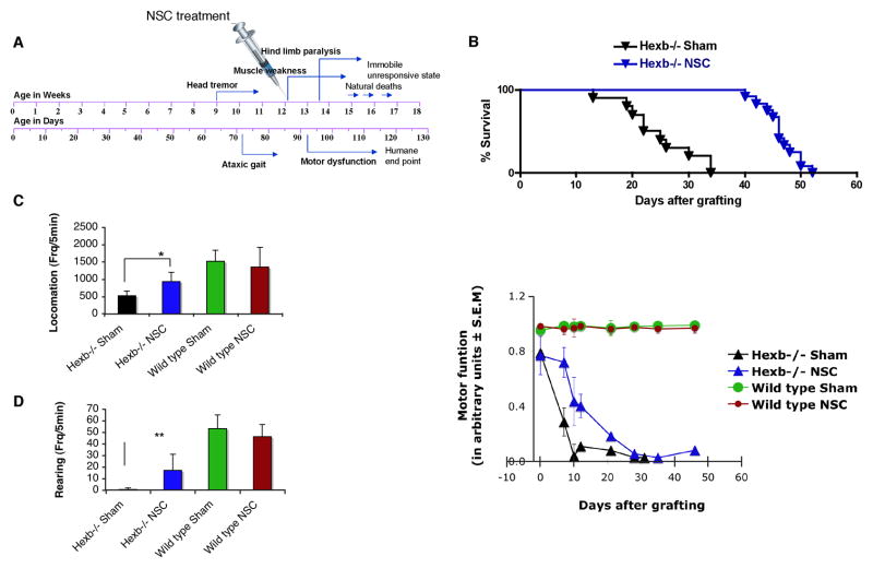
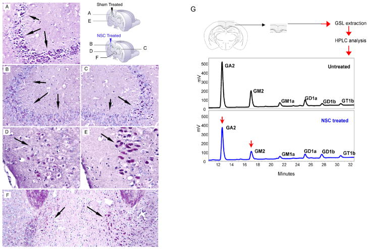
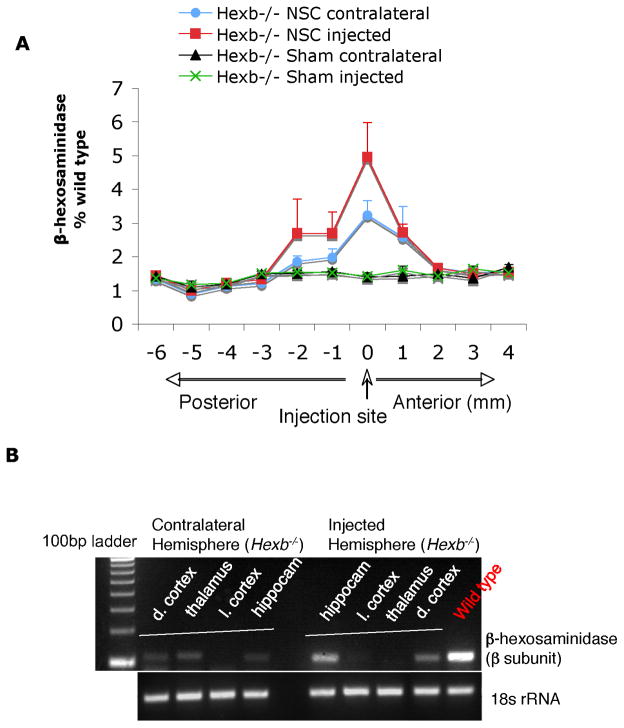
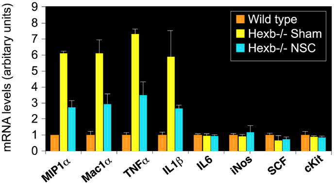
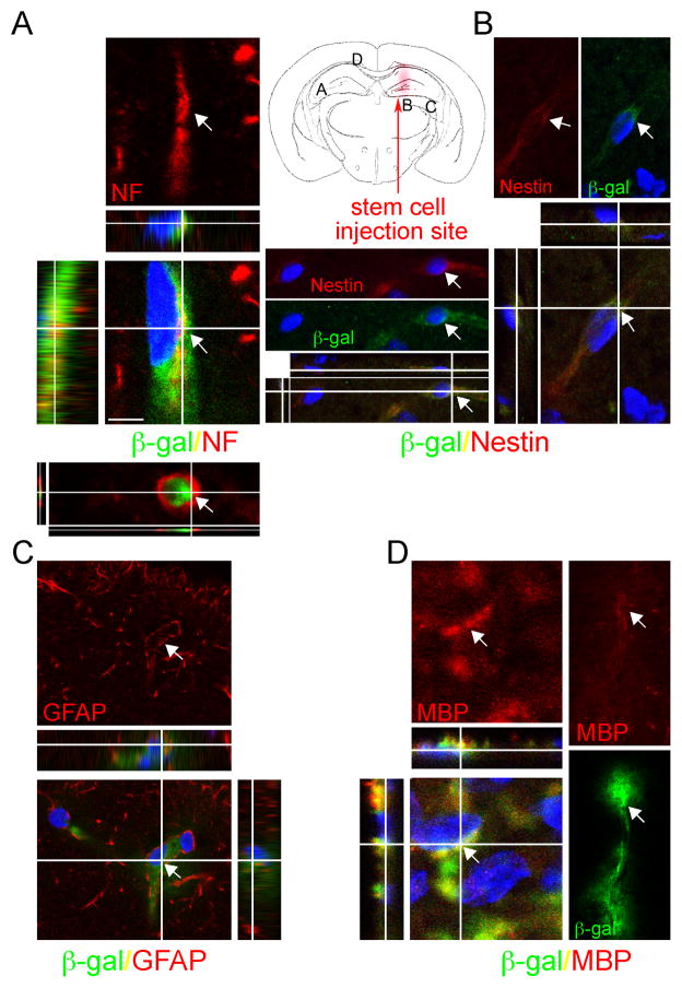
Although we and others have demonstrated that neural stem cells (NSCs) may impact such neurogenetic conditions as lysosomal storage diseases when transplanted at birth, it has remained unclear whether such interventions can impact well-established mid-stage disease, a situation often encountered clinically. Here we report that when NSCs were injected intracranially into the brain of adult symptomatic Sandhoff (Hexb(-/-)) mice, cells migrated far from the injection site and integrated into the host cytoarchitecture, restoring beta-hexosaminidase enzyme activity and promoting neuropathologic and behavioral improvement. Mouse lifespan increased, neurological function improved, and disease progression was slowed. These clinical benefits correlated with neuropathological correction at the cellular and molecular levels, reflecting the multiple potential beneficial actions of stem cells, including enzyme cross-correction, cell replacement, tropic support, and direct anti-inflammatory action. Pathotropism (i.e., migration and homing of NSCs to pathological sites) could be imaged in real time by magnetic resonance imaging. Differentially expressed chemokines might play a role in directing the migration of transplanted stem cells to sites of pathology. Significantly, the therapeutic impact of NSCs implanted in even a single location was surprisingly widespread due to both cell migration and enzyme diffusion. Because many of the beneficial actions of NSCs observed in newborn brains were recapitulated in adult brains to the benefit of Sandhoff recipients, NSC-based interventions may also be useful in symptomatic subjects with established disease.


Figures

Figure 1
Figure 1
I. NSC transplants survive and engraft in Sandhoff disease Hexb−/− mouse model. Adult symptomatic mice 12–13 weeks of age were used. Using a stereotaxic frame (KOPF®), through a 0.9-mm diameter hole in the skull (2-mm posterior to bregma; 1.5-mm lateral to sagittal suture), 4 μl (8 × 10⁴ cells/μl) of C17.2 cells in PBS was injected unilaterally into the hippocampus (right hemisphere) under isofluorane anaesthesia. Mock-grafted controls received the same volume of PBS. Coronal brain sections were permeabilized with detergents and exposed to a chromogenic substrate X-gal to detect β-gal-labeled donor cells. The blue cells (X-gal histochemical reaction, arrows) have engrafted in the recipient brain with widespread migration, distribution, and integration. Representative images of the brain (A–C) from 15-week-old Hexb−/− mice are shown (n=7). Schematic diagram (a–c) illustrates where images are from (images A–C, respectively) in relation to injection site. Scale bar = 10 μm. Counter stain = neutral red. II. Noninvasive MRI tracking of neural stem cells in live mice. T2-weighted image acquired with a fast spin-echo sequence (TR=3s, TE=48ms), 1-mm coronal slice with an in-plane resolution of 234 μm, at posttransplantation day 38. Injection site of Sinerem®-labeled stem cells is clearly visible (red arrow) in the right hemisphere of Hexb−/− mice, while minimal change in signal intensity is evident in the wild-type transplanted mice which had received the same number of Sinerem®-labeled stem cells. More NSCs (black, red arrow) are located juxtaposed to T2 hyperintensity in the cortex (yellow arrow, which could represent increased inflammation) compared to similarly transplanted NSCs in wild type mice, which show no such T2 signal (yellow arrow) & very few, if any, NSCs (black, red arrow) accumulating in the cortex). Although the main migration streams were detected with MR, the technique does not have the sensitivity to detect lower cell densities readily detected histologically. n=2 per group.
Figure 2
Figure 2. NSC transplants improve lifespan and delay rate of disease progression in symptomatic adult Hexb−/− mice
NSCs, not engineered to overexpress HexB, were injected into one site of non-immunosuppressed adult SD mice. (A) Schematic of symptom onset and kinetics of disease progression in Sandhoff disease (Hexb−/−) mice. (B) Survival of NSC-transplanted mice (n=12) compared with sham-treated control Hexb−/− mice (n=10). NSC transplantation prolonged Hexb−/− mice lifespan by 21 days (p<0.0001, Log rank test), resulting in a 19% improvement in survival. (C, D) Neurologic function scored in an automated activity monitor, which records infrared beam breaks in the X -Y plane (locomotion) and vertical (rearing) movements (data at 2 weeks posttreatment, 15 weeks of age). Data represent mean ± s.e.m, n=6 per group. An unpaired two-tailed Student’s t-test was used to compare data for significance. (E) Temporal profile of motor function before and after NSC treatment. Motor function was assessed using a horizontal bar-crossing test measuring the latency to cross or fall from the bar. Motor-function decline was significantly delayed in the NSC-treated SD mice, compared to the sham-treated SD control group (p<0.0001, two-way ANOVA). Data represent mean ± s.e.m, n=7–12.
Figure 3
Figure 3. Reduction of GSL storage in brains of NSC-treated Hexb−/− mice
(A–F) Representative PAS staining from brain sections of NSC-treated and sham-treated control mice at 15 weeks of age (n=4 per group). Storage GSLs were visualised in frozen brain sections by PAS staining (red). Panels A and E show sham-injected hemisphere of control Hexb−/− mice; panels B and D represent injected hemisphere of NSC-treated Hexb−/− mice; panel C represents noninjected hemisphere of NSC-treated Hexb−/− mice; panel F represents a region of thalamus of NSC-treated Hexb−/− mice. Hippocampus CA3 region (A–C), mediodorsal thalamic nucleus (F), and late entrohinal cortex (D, F) are shown. By 3 weeks posttransplant, the hippocampus of NSC-treated Hexb−/− mice showed less PAS-positive GSL storage (B and C, compared with A), the most significant reduction achieved in the injected hemisphere, compared to the noninjected hemisphere in the same animal (C compared with B). (G) Representative HPLC profile of cerebral cortical region of NSC-treated and sham-treated Hexb−/− mice are shown 2 weeks posttreatment (n=4 per group). GM2 was reduced by 19±1% (p=0.004) while GA2 was reduced by 8±0.3% (p=0.015) in the brain region shown, as analyzed by HPLC. An unpaired two-tailed Student’s t-test was used to compare data for significance.
Figure 4
Figure 4. Increased β-hexosaminidase enzyme levels in brains of NSC-treated Hexb−/− mice
(A) Representative Hex levels in serial coronal brain sections of individual hemisphere from NSC-treated and sham-treated Hexb−/− mice at 4 weeks posttransplant. Brain homogenates were made from 12 individual brain slices, as per schematic diagram, and hex levels were measured against 4-MUGlcNAc artificial substrate (mean ± s.e.m., n=3). Ipsilateral represents the injected hemisphere, while contralateral represents the noninjected hemisphere. (B) RT-PCR analysis for β-subunit expression of Hex from local brain regions. Two or more brain regions (dorsal cortex/thalamus/lateral cortex/hippocampus) expressed the Hexβ-subunit gene in each hemisphere, indicative of donor-derived cell migration and brain engraftment upon unilateral (hippocampus) NSC transplantation.
Figure 5
Figure 5. Reduced inflammatory marker expression in brains of NSC-treated Hexb−/− mice
Expression of inflammatory genes in mouse brains as measured by RT-PCR. The relative gene expression normalized with 18S RNA (arbitrary units) is shown on the graphs. Accompanied by increased levels of microglial/macrophage activation marker Mac1α (Cd11b) (p<0.001), proinflammatory cytokines TNFα and IL-1β and the chemokine MIP1α (CCL3), expression levels were high in the diseased mice, compared to the wild-type mice (p<0.001). Upon NSC treatment, all upregulated genes were reduced significantly when the age-matched sham-treated control group was compared (p<0.001). No significant changes in IL-6, iNOS, SCF or c-kit expression were observed. Data are mean ± s.e.m. based on 3–5 animals in each group (R and L hemispheres averaged together). An unpaired two-tailed Student’s t-test was used to compare data for significance.
Figure 6
Figure 6. Engrafted NSCs differentiate into a range of neural cell types in the Hexb−/− mouse brain
Following NSC transplantation at 13 weeks of age, brains of Hexb−/− mice were processed for cell type characterization using immunoflourescence staining for both LacZ expressed β-gal and neural cell-type specific markers. At 3 weeks posttransplant, NSCs stably engrafted in the Hexb−/− mouse brain, differentiating into a range of neural cell types. (A–D) Representative donor-derived cells, recognized by their immunoreactivity to β-gal (green, bottom), coexpress neuronal markers such as neurofilament (NF), astroglial marker GFAP, oligodentrocyte marker MBP and nestin, a marker of undifferentiated neural precursors. Merged images show blue DAPI nuclear staining identifies all cells in the field (bottom). Each merged image is also shown as an orthogonal projection composed of 9–16 optical Z-planes, 0.5–1 μm thick. Arrows in each panel of a given column indicate the same cell. Scale bar = 10 μm. (see also Supplementary Fig 2).

References

    1. Wraith JE. Lysosomal disorders. Semin Neonatol. 2002;7:75–83. - PubMed
    1. Wada R, Tifft CJ, Proia RL. Microglial activation precedes acute neurodegeneration in Sandhoff disease and is suppressed by bone marrow transplantation. Proc Natl Acad Sci U S A. 2000;97:10954–9. - PMC - PubMed
    1. Myerowitz R, Lawson D, Mizukami H, Mi Y, Tifft CJ, Proia RL. Molecular pathophysiology in Tay-Sachs and Sandhoff diseases as revealed by gene expression profiling. Hum Mol Genet. 2002;11:1343–50. - PubMed
    1. Jeyakumar M, Thomas R, Elliot-Smith E, Smith DA, van der Spoel AC, d’Azzo A, Perry VH, Butters TD, Dwek RA, Platt FM. Central nervous system inflammation is a hallmark of pathogenesis in mouse models of GM1 and GM2 gangliosidosis. Brain. 2003;126:974–87. - PubMed
    1. Sango K, Yamanaka S, Hoffmann A, Okuda Y, Grinberg A, Westphal H, McDonald MP, Crawley JN, Sandhoff K, Suzuki K, et al. Mouse models of Tay-Sachs and Sandhoff diseases differ in neurologic phenotype and ganglioside metabolism. Nat Genet. 1995;11:170–6. - PubMed

Publication types