

On the biomechanics of vaginal birth and common sequelae
- PMID: 19591614
- PMCID: PMC2897058
- DOI: 10.1146/annurev-bioeng-061008-124823
On the biomechanics of vaginal birth and common sequelae
Abstract
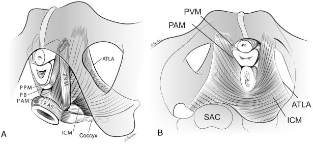
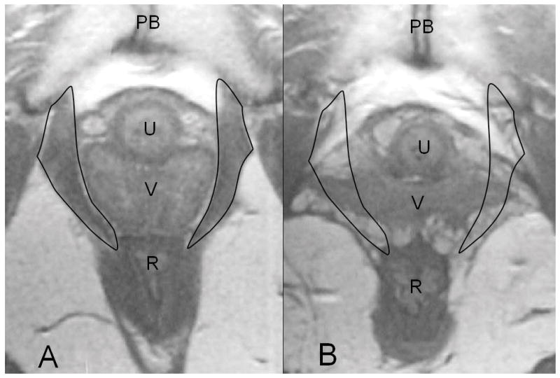
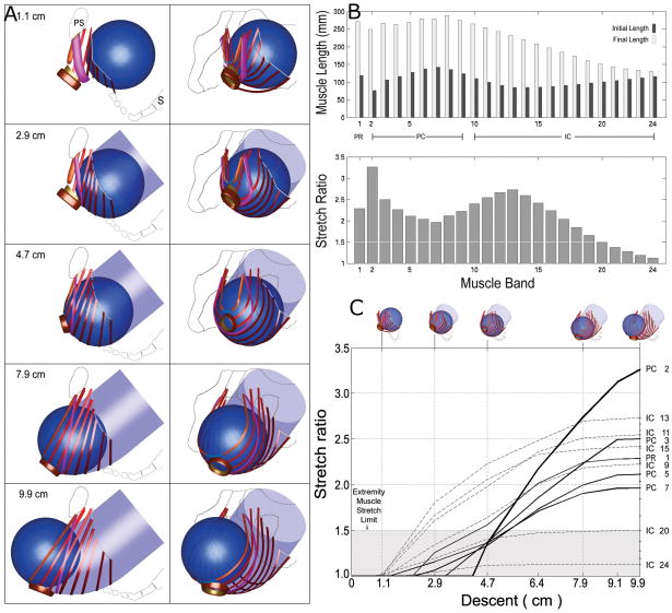
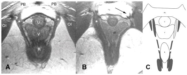
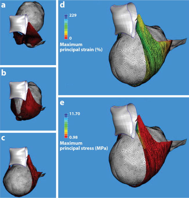
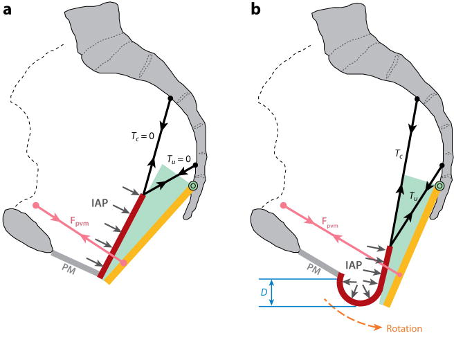
Approximately 11% of U.S. women undergo surgery for pelvic floor dysfunction, including genital organ prolapse and urinary and fecal incontinence. The major risk factor for developing these conditions is giving vaginal birth. Vaginal birth is a remarkable event about which little is known from a biomechanical perspective. We first review the functional anatomy of the female pelvic floor, the normal loads acting on the pelvic floor in activities of daily living, and the functional capacity of the pelvic floor muscles. Computer models show that the stretch ratio in the pelvic floor muscles can reach an extraordinary 3.26 by the end of the second stage of labor. Magnetic resonance images provide evidence that show that the pelvic floor regions experiencing the most stretch are at the greatest risk for injury, especially in forceps deliveries. A conceptual model suggests how these injuries may lead to the most common form of pelvic organ prolapse, a cystocele.
Figures
References
-
- Mant J, Painter R, Vessey M. Epidemiology of genital prolapse: observations from the Oxford Planning Association Study. Brit J Obstet Gynaecol. 1997;104:579–85. - PubMed
-
- Hodges PW, Sapsford R, Pengel LH. Postural and respiratory functions of the pelvic floor muscles. Neurourol Urodyn. 2007;26:362–71. - PubMed
-
- Morgan DM, Kaur G, Hsu Y, Fenner DE, Guire K, et al. Does vaginal closure force differ in the supine and standing positions? Am J Obstet Gynecol. 2005;192:1722–28. - PubMed
-
- Baragi R, DeLancey JOL, Caspari R, Howard DH, Ashton-Miller JA. Differences in pelvic floor area between African American and European American Women. Am J Obstet Gynecol. 2002;187:111–15. - PubMed
Publication types
MeSH terms
Grants and funding
LinkOut - more resources
Full Text Sources
Medical
