. 2009 Dec;78(3):413-24.
doi: 10.1016/j.mvr.2009.06.009. Epub 2009 Jul 8.

Reproductive age-related changes in the blood brain barrier: expression of IgG and tight junction proteins

Affiliations

Reproductive age-related changes in the blood brain barrier: expression of IgG and tight junction proteins

Shameena Bake et al. Microvasc Res. 2009 Dec.

Abstract

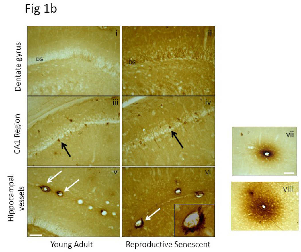
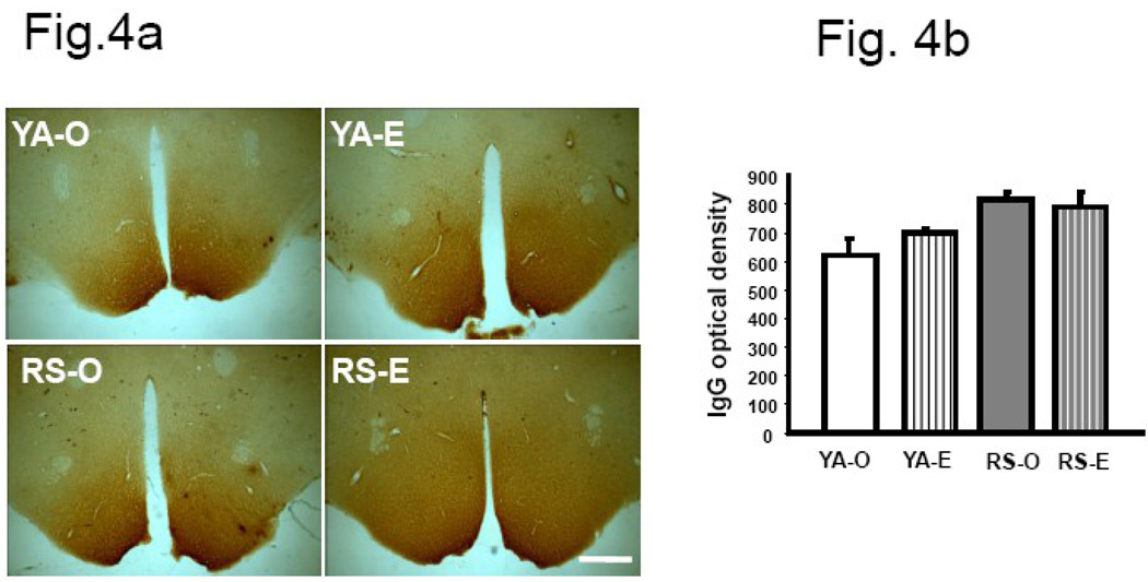
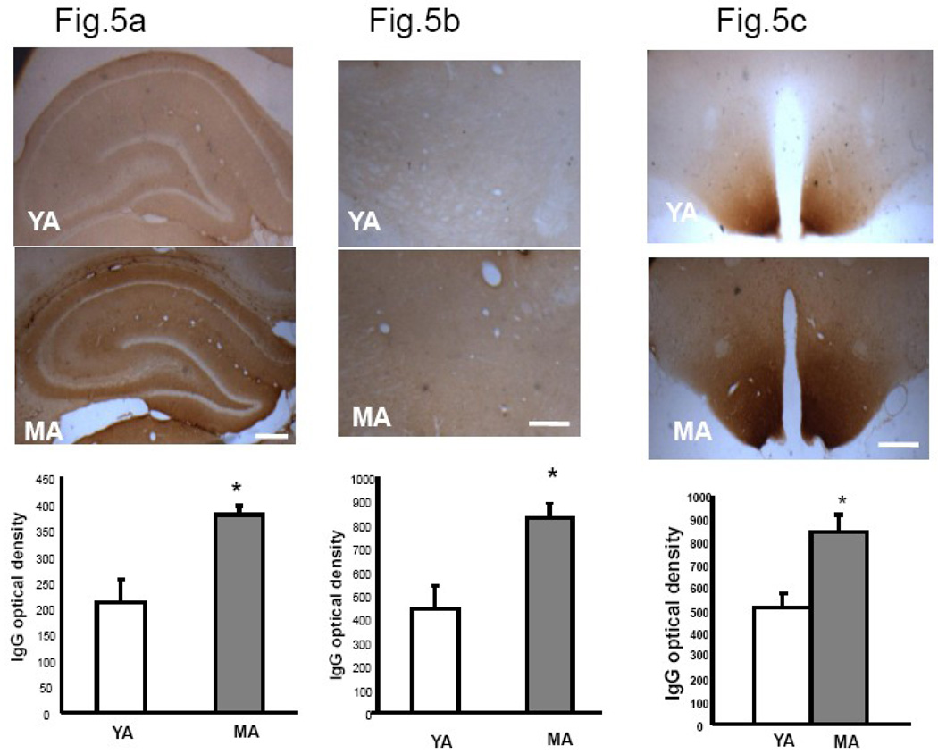
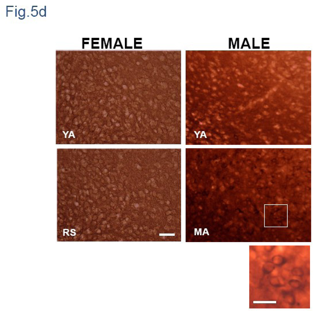
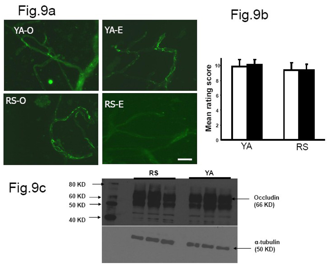
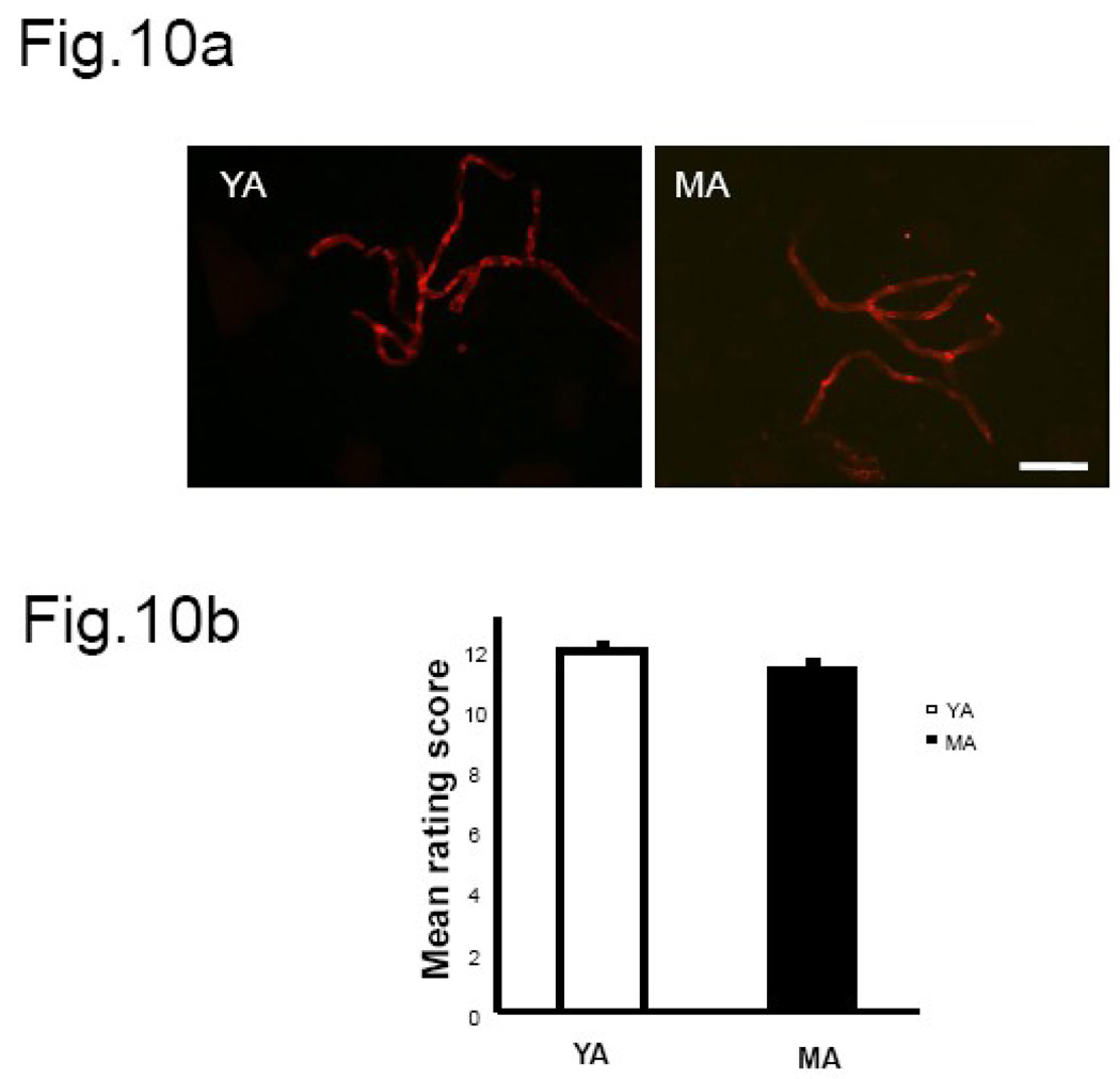
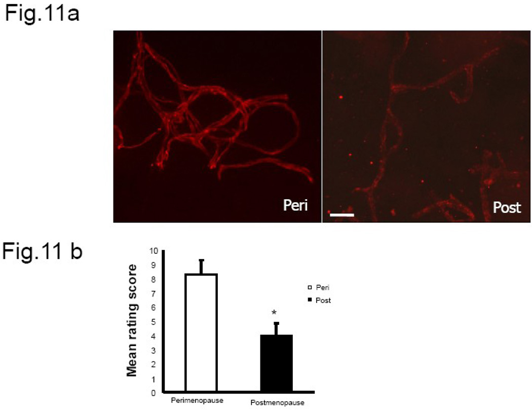
We previously demonstrated that there is a significantly greater transfer of intravenously-injected Evan's blue dye into the forebrain of acyclic (reproductive senescent) females compared to young adult females, indicating that blood brain barrier permeability is compromised in the reproductive senescent forebrain. The present study examined brain IgG expression and microvessel tight junction proteins to assess ovarian age-related changes in microvascular permeability, and further compared young and senescent females with age-matched males to distinguish changes attributable to age and reproductive senescence. Blood brain barrier breakdown are often associated with increased extravasation of plasma proteins and high levels of immunoglobulin G (IgG) in brain. In the present study, IgG expression was dramatically increased in the hippocampus and thalamus, but not the hypothalamus of reproductive senescent females compared to young adult females. In males, IgG expression was increased in all these regions in middle-aged animals (aged-matched to senescent females) as compared to young males (age-matched to the young adult females). Furthermore, the proportion of hippocampal microvessels with perivascular IgG immunoreactivity was significantly greater in reproductive senescent females as compared to young adult females, while middle-aged males and young adult males did not differ. The tight junctions between adjacent microvascular endothelial cells regulated by transmembrane proteins such as claudin-5 and occludin play a critical role in maintaining the blood brain barrier integrity. Increased hippocampal IgG expression in senescent females was paralleled by poor junctional localization of the tight junction protein claudin-5 in hippocampal microvessels. However, there was no difference in hippocampal claudin-5 localization between young adult and middle-aged males, indicating that dysregulation of this junctional protein was associated with ovarian aging. Parallel studies in human brain microvessels also revealed age-dependent disruption in claudin-5 distribution in post-menopausal women compared to pre-menopausal women. Collectively, these data support the hypothesis that constitutive loss of barrier integrity in the forebrain during reproductive senescence may be due, in part, to the selective loss of tight junction proteins in endothelial junctions.


Figures

Fig.1
Fig.1. IgG expression in female and male brain
(a) Coronal brain sections from young adult and reproductive senescent females as well as age-matched males were processed for IgG immunoreactivity. Visual inspection indicated overall greater IgG immunoreactive product in the forebrain of older males and females compared to their younger counterparts. Regional intensity of IgG staining was quantitated by densitometry from digitized images. (b) IgG expression was seen in the parenchyma (i and ii), in neurons (iii and iv) and blood vessels (v and vi) throughout the brain and images shown here are from the hippocampal formation. In microvessels, IgG-positive immunoreactivity was seen as a halo surrounding the lumen of blood vessels (vii and viii) Bars 1b i–vi: 50µm, vii–viii: 20µm.
Fig.2
Fig.2. IgG immunoreactivity in the hippocampus of the young adult and reproductive senescent brain
Coronal sections of the young adult and reproductive senescent brain containing the dorsal hippocampus were scanned and the intensity of IgG staining in this region was quantified from the digitized images using Quantity One® software (Bio-Rad, CA). (a) IgG immunoreactivity was stronger in the hippocampus of senescent females as compared to young adults and mean (±SEM) intensity of IgG immunoreactivity is shown in the histogram in (b). Histogram indicates mean (±SEM) optical density of IgG immunoreactivity in hippocampus of young and senescent animals. No significant estrogen effect was observed in either age group. (N=4 per group, * p<0.05) Bar =50 µm. Key: YA: Young adult, RS: reproductive senescent, O: ovariectomized and replaced with control pellet, E: ovariectomized and replaced with estrogen pellet.
Fig.3
Fig.3. IgG immunoreactivity in the thalamus of the young adult and reproductive senescent brain
(a) Stronger IgG immunoreactivity was seen in the thalamus of reproductive senescent females as compared to those of young adult females. (3b) Histogram indicates mean (±SEM) optical density of IgG immunoreactivity in thalamus of young and senescent animals. No significant estrogen effect was observed in either age group. N=4 per group, * p<0.05. Bar=50 µm. Key: YA: Young adult, RS: reproductive senescent, O: ovariectomized and replaced with control pellet, E: ovariectomized and replaced with estrogen pellet.
Fig.4
Fig.4. IgG immunoreactivity in the hypothalamus of the young adult and reproductive senescent brain
(a) Although strong IgG immunoreactivity was visible in the hypothalamus of young and senescent females, there was no difference in staining intensity due to age or estrogen treatment. (b) Histogram shows mean (±SEM) IgG optical density. N=4 per group. Bar=50 µm. Key: YA: young adult, RS: reproductive senescent, O: ovariectomized and replaced with control pellet, E: ovariectomized and replaced with estrogen pellet.
Fig.5
Fig.5. IgG immunoreactivity in hippocampus, thalamus and hypothalamus of young adult and middle-aged males
Coronal sections of young adult and middle-aged males were scanned and optical density was quantified from digitized images using Quantity One® software (Bio-Rad, CA). Histograms represent mean (±SEM) IgG optical density. Stronger immunoreactivity was seen in hippocampus (6a) thalamus (6b) and hypothalamus (6c) of middle-aged males compared to young adults. (6d) Higher magnification view of the hypothalamus shows IgG labeling was largely restricted to the neuropil of females, while in males, intensely stained neuropil as well as prominent cellular labeling was visible. Inset is a magnified image of the hypothalamus of the middle aged male indicating cellular IgG label. YA: Young adult, RS: reproductive senescent (female) MA: Middle-Aged male (age matched to RS female). Bar=50 µm; Inset Bar= 25 µm
Fig.6
Fig.6. Cellular characterization of microvessels
Microvessels were stained with antibodies specific for the endothelial marker, Pecam-1 (in red) and the glial marker, GFAP (in green). The vessels were also counterstained with nuclear dye, Hoeschst 33342 (in blue). Co-localization of pecam-1 and GFAP was observed at several regions of the individual vessel (indicated in white arrows).
Fig.7
Fig.7. Immunostaining for tight junction proteins
Antibodies specific for (a) occludin and (b) claudin-5 were used for immunohistochemical detection of tight junction proteins in microvessels. These antibodies recognized size-appropriate bands in Western blots. (c) Shown here is an isolated hippocampal microvessel from an estrogen-treated young adult female probed with the claudin-5 antibody. Arrows indicate bright, continuous junctional localization of this protein. Bar=50 µm.
Fig.8
Fig.8. Claudin-5 immunofluorescence in hippocampal microvessels
(a) Claudin-5 was expressed in hippocampal microvessels derived from both young adult and reproductive senescent females, however claudin-5 expression was brighter and more junctional in microvessels from young adults as compared to those from reproductive senescent females. (b) Histogram represents the average (±SEM) composite rating scores for continuity and junctional localization for claudin-5. Estrogen treatment (closed bars) did not affect the ratings scores in either young adult or senescent females. (c) Western blot analysis showed no change in claudin-5 expression between young adults and reproductive senescent females. N=4–5 per group, 6 vessels were rated per animal, * P<0.05. Key: YA: young adult, RS: reproductive senescent, O: ovariectomized and replaced with control pellet, E: ovariectomized and replaced with estrogen pellet. Bar=50 µm.
Fig.9
Fig.9. Occludin immunofluorescence in hippocampal microvessels
(a) Occludin staining was brighter and well-localized to the cell junctions in microvessels derived from both young adult females and reproductive senescent animals. (b) Histogram represents the average (±SEM) composite rating score for continuity and junctional localization for occludin. There was no difference in occludin immunostaining in microvessel tight junctions of young adults and reproductive senescent animals. (c) No change in occludin expression was seen in Western blots from young adult and reproductive senescent microvessels. N=4–5 per group, 6 vessels were rated per animal. Key: YA: young adult, RS: reproductive senescent, O: ovariectomized and replaced with control pellet, E: ovariectomized and replaced with estrogen pellet. Bar=50 µm.
Fig.10
Fig.10. Claudin-5 immunofluorescence in hippocampal microvessels from male rats
(a) Claudin-5 staining was bright and well-localized to the cell junctions in microvessels derived from both 3 month old and middle-aged males. (b) Histogram represents the average (±SEM) composite rating score for continuity and junctional localization for claudin-5. There was no difference in claudin-5 expression pattern between young and middle-aged males. N=4 per group, 6 vessels were rated per animal, * P<0.05. Key: YA: young adult, MA: middle aged. Bar=50 µm.
Fig.11
Fig.11. Claudin-5 immunofluorescence in microvessels from human brain
(a) Claudin-5 staining in human brain microvessels indicates a change in expression pattern between the pre-menopausal and post-menopausal group. Microvessels collected from younger women showed continuous junctional distribution of claudin-5 compared to older subjects. (b) Histogram represents the average (±SEM) composite rating score for continuity and junctional localization for claudin-5. N= 3–4 per group, 6–8 vessels were rated per sample, * P<0.05. Key: Bar=50 µm.

References

    1. Aberdeen GW, et al. Vascular endothelial growth factor mediates the estrogen-induced breakdown of tight junctions between and increase in proliferation of microvessel endothelial cells in the baboon endometrium. Endocrinology. 2008;149:6076–6083. - PMC - PubMed
    1. Aihara N, et al. Immunocytochemical localization of immunoglobulins in the rat brain: relationship to the blood-brain barrier. J Comp Neurol. 1994;342:481–496. - PubMed
    1. Andoh T, Kuraishi Y. Direct action of immunoglobulin G on primary sensory neurons through Fc gamma receptor I. FASEB J. 2004;18:182–184. - PubMed
    1. Andreeva AY, et al. Protein Kinase C Regulates the Phosphorylation and Cellular Localization of Occludin. J Biol Chem. 2001;276:38480–38486. - PubMed
    1. Arai K, et al. Deterioration of spatial learning performances in lipopolysaccharide-treated mice. J Pharmacol. 2001;87:195–221. - PubMed

Publication types

MeSH terms