. 2009 Sep;30(5):772-6.
doi: 10.1016/j.neuro.2009.06.011. Epub 2009 Jul 8.

Primary brain targets of nerve agents: the role of the amygdala in comparison to the hippocampus

Affiliations

Primary brain targets of nerve agents: the role of the amygdala in comparison to the hippocampus

Vassiliki Aroniadou-Anderjaska et al. Neurotoxicology. 2009 Sep.

Abstract

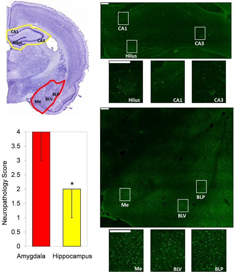
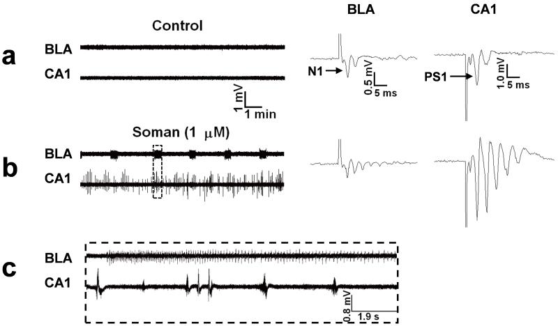
Exposure to nerve agents and other organophosphorus acetylcholinesterases used in industry and agriculture can cause death, or brain damage, producing long-term cognitive and behavioral deficits. Brain damage is primarily caused by the intense seizure activity induced by these agents. Identifying the brain regions that respond most intensely to nerve agents, in terms of generating and spreading seizure activity, along with knowledge of the physiology and biochemistry of these regions, can facilitate the development of pharmacological treatments that will effectively control seizures even if administered when seizures are well underway. Here, we contrast the pathological (neuronal damage) and pathophysiological (neuronal activity) findings of responses to nerve agents in the amygdala and the hippocampus, the two brain structures that play a central role in the generation and spread of seizures. The evidence so far suggests that exposure to nerve agents causes significantly more damage in the amygdala than in the hippocampus. Furthermore, in in vitro brain slices, the amygdala generates prolonged, seizure-like neuronal discharges in response to the nerve agent soman, at a time when the hippocampus generates only interictal-like activity. In vivo experiments are now required to confirm the primary role that the amygdala seems to play in nerve agent-induced seizure generation.


Figures

Figure 1
Figure 1
Neuronal degeneration in the amygdala is greater than in the hippocampus, 24 hours after status epilepticus induced by injection of 1.4×LD50 soman (154 μg/kg BW), in rats. Photomicrographs of Cresyl Violet (left) and FluoroJade-C staining (right) from one representative animal. The Cresyl Violet photomicrograph outlines the brain regions (amygdala in red; hippocampus in yellow) and indicates the hippocampal subfields (CA1, CA3 and hilus) and amygdala nuclei (Me, Medial; BLV, basolateral ventral; BLP, basolateral posterior) which are shown in the FluoroJade-C sections (magnification 200×). The bar graph shows the neuropathology score in the amygdala and the hippocampus (Median ± Range; group data from 6 animals). The score assessment was done using a qualitative scale (see McDonough et al., 1995 and Myhrer et al., 2005): 0 = no damage; 1 = 1 to 10% minimal damage; 2 = 11 to 25% mild damage; 3 = 26 to 45% moderate damage; 4 > 45% severe damage. The final score was the average from 5 successive coronal brain sections from each animal. Scale bars are 200 μm. *p<0.05 using Wilcoxon Signed Ranks Test to compare the two brain regions.
Figure 2
Figure 2. Soman induces ictal-like activity in the amygdala and interictal-like activity in the hippocampus
Extracellular field recordings, in gap-free mode, were simultaneously obtained in the BLA and the stratum pyramidale of the CA1 hippocampal area, in slices containing both regions. Stimulus pulses were applied every 30 sec to sample the evoked field potentials. Note the amplitude scales of the field potentials; field potentials in the amygdala have substantially smaller amplitude than hippocampal field potentials due to the structure of the amygdala network which does not favor generation of strong dipoles. (a) Field potentials in the BLA, evoked by stimulation of the external capsule, consisted of one major negative component (N1), followed by one or more lower-amplitude, late components. In the CA1 area, field potentials evoked by stimulation of the Schaffer collaterals consisted of a large population spike (PS1), which was often followed by one or two low-amplitude, negative components. No spontaneous activity was present in the BLA or the CA1 area (left panel). (b) Exposure to 1 μM soman for 30 min induced spontaneous, prolonged episodes of synchronous neuronal discharges resembling brain seizures, in the BLA. In the CA1 area, soman exposure produced additional population spikes in the enhanced evoked response, as well as spontaneous, interictal-like bursts. (c) A prolonged ictal-like discharge from the BLA and interictal-like activity from the hippocampus on an expanded time scale (taken from the rectangle-outline shown in (b)). In no case did the hippocampus display seizure-like activity (see Apland et al., 2009). The effects of soman were not reversible; epileptiform activity was maintained after washing out soman and throughout the recording period (for more than 3 hours).

References

    1. Akaike K, Tanaka S, Hideshi Tojo H, Fukumoto S, Imamura S, Takigawa M. Kainic acid-induced dorsal and ventral hippocampal seizures in rats. Brain Res. 2001;900:65–71. - PubMed
    1. Apland JP, Aroniadou-Anderjaska V, Braga MF. Soman induces ictogenesis in the amygdala and interictal activity in the hippocampus that are blocked by a GluR5 kainate receptor antagonist in vitro. Neuroscience. 2009;159:380–9. - PMC - PubMed
    1. Aroniadou-Anderjaska V, Qashu F, Braga MF. Mechanisms regulating GABAergic inhibitory transmission in the basolateral amygdala: Implications for epilepsy and anxiety disorders. Amino Acids. 2007;32:305–15. - PubMed
    1. Aroniadou-Anderjaska V, Fritsch B, Qashu F, Braga MF. Pathology and pathophysiology of the amygdala in epileptogenesis and epilepsy. Epilepsy Res. 2008;78:102–116. - PMC - PubMed
    1. Avoli M. Do interictal discharges promote or control seizures? Experimental evidence from an in vitro model of epileptiform discharge. Epilepsia. 2001;42:2–4. - PubMed

Publication types