

Transplantation of adrenal cortical progenitor cells enriched by Nile red
- PMID: 19592014
- PMCID: PMC2749914
- DOI: 10.1016/j.jss.2009.04.021
Transplantation of adrenal cortical progenitor cells enriched by Nile red
Abstract
Background: The adrenal cortex may contain progenitor cells useful for tissue regeneration. Currently there are no established methods to isolate these cells.
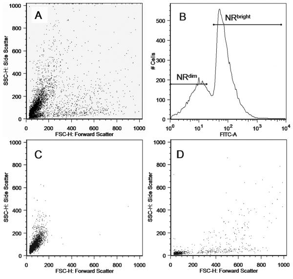
Material and methods: Murine adrenal cells were sorted into a Nile-red-bright (NR(bright)) and a Nile-red-dim (NR(dim)) population of cells according to their degree of cholesterol content revealed by Nile red fluorescence. The cells were transplanted under the renal capsule to determine their ability for regeneration.
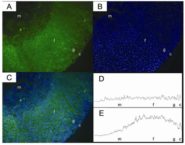
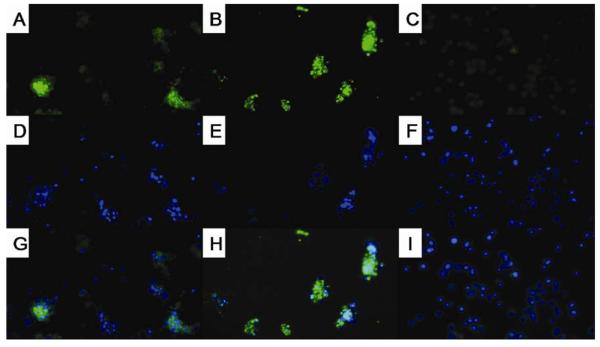
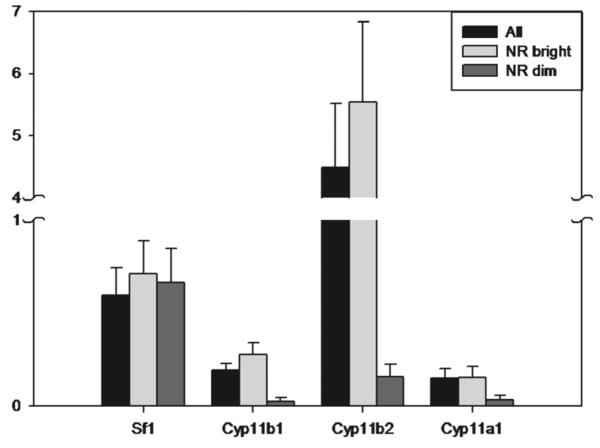
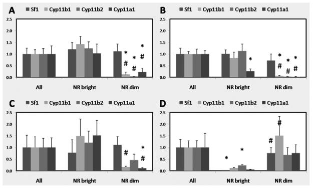
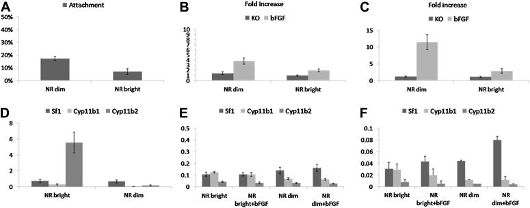
Results: The NR(bright) cells contained an abundance of lipid droplets, whereas the NR(dim) cells contained little. The NR(bright) cells expressed Sf1 and the more differentiated adrenal cortical genes, including Cyp11a1, Cyp11b1, and Cyp11b2, whereas the NR(dim) cells expressed Sf1 but not the more differentiated adrenal cortical genes. After 56 d of implantation in unilateral adrenalectomized mice, the NR(dim) cells expressed Sf1 and the more differentiated adrenal cortical genes, whereas the NR(bright) cells ceased to express Sf1 as well as the more differentiated adrenal cortical genes. NR(dim) cells also proliferated in the presence of basic fibroblast growth factor.
Conclusions: The population of NR(dim) cells contained adrenal cortical progenitor cells that can proliferate and give rise to differentiated daughter cells. These cells may be useful for adrenal cortical regeneration.
Figures
Comment in
-
Nile red staining helps select cells with adrenocortical progenitor cell-like phenotype.J Surg Res. 2010 Jun 1;161(1):34-5. doi: 10.1016/j.jss.2009.07.019. Epub 2009 Aug 12. J Surg Res. 2010. PMID: 20031173 No abstract available.
Similar articles
-
Nile red staining helps select cells with adrenocortical progenitor cell-like phenotype.J Surg Res. 2010 Jun 1;161(1):34-5. doi: 10.1016/j.jss.2009.07.019. Epub 2009 Aug 12. J Surg Res. 2010. PMID: 20031173 No abstract available.
-
Adrenal cortical cell transplantation.J Pediatr Surg. 2004 Dec;39(12):1856-8. doi: 10.1016/j.jpedsurg.2004.08.006. J Pediatr Surg. 2004. PMID: 15616950
-
Basic fibroblast growth factor delivery enhances adrenal cortical cellular regeneration.Tissue Eng Part A. 2009 Aug;15(8):2093-101. doi: 10.1089/ten.tea.2008.0305. Tissue Eng Part A. 2009. PMID: 19196135 Free PMC article.
-
How to use Nile Red, a selective fluorescent stain for microalgal neutral lipids.J Microbiol Methods. 2016 Sep;128:74-79. doi: 10.1016/j.mimet.2016.07.011. Epub 2016 Jul 16. J Microbiol Methods. 2016. PMID: 27432343 Review.
-
Adrenal cortical and chromaffin stem cells: Is there a common progeny related to stress adaptation?Mol Cell Endocrinol. 2017 Feb 5;441:156-163. doi: 10.1016/j.mce.2016.09.011. Epub 2016 Sep 13. Mol Cell Endocrinol. 2017. PMID: 27637345 Review.
Cited by
-
[Gene and cell therapy of adrenal pathology: achievements and prospects].Probl Endokrinol (Mosk). 2021 Dec 2;67(6):80-89. doi: 10.14341/probl12818. Probl Endokrinol (Mosk). 2021. PMID: 35018764 Free PMC article. Review. Russian.
-
Expression of progenitor markers is associated with the functionality of a bioartificial adrenal cortex.PLoS One. 2018 Mar 29;13(3):e0194643. doi: 10.1371/journal.pone.0194643. eCollection 2018. PLoS One. 2018. PMID: 29596439 Free PMC article.
-
Adrenocortical cell transplantation reverses a murine model of adrenal failure.J Pediatr Surg. 2011 Jun;46(6):1208-13. doi: 10.1016/j.jpedsurg.2011.03.057. J Pediatr Surg. 2011. PMID: 21683224 Free PMC article.
-
KIAA0101 is overexpressed, and promotes growth and invasion in adrenal cancer.PLoS One. 2011;6(11):e26866. doi: 10.1371/journal.pone.0026866. Epub 2011 Nov 11. PLoS One. 2011. PMID: 22096502 Free PMC article.
-
Label-free enrichment of adrenal cortical progenitor cells using inertial microfluidics.PLoS One. 2012;7(10):e46550. doi: 10.1371/journal.pone.0046550. Epub 2012 Oct 4. PLoS One. 2012. PMID: 23056341 Free PMC article.
References
-
- Iannaccone P, Morley S, Skimina T, Mullins J, Landini G. Cord-like mosaic patches in the adrenal cortex are fractal: implications for growth and development. Faseb J. 2003;17:41–43. - PubMed
-
- Weinberg WC, Howard JC, Iannaccone PM. Histological demonstration of mosaicism in a series of chimeric rats produced between congenic strains. Science. 1985;227:524–527. - PubMed
-
- Ogishima T, Suzuki H, Hata J, Mitani F, Ishimura Y. Zone-specific expression of aldosterone synthase cytochrome P-450 and cytochrome P-45011 beta in rat adrenal cortex: histochemical basis for the functional zonation. Endocrinology. 1992;130:2971–2977. - PubMed
-
- Mitani F, Suzuki H, Hata J, Ogishima T, Shimada H, Ishimura Y. A novel cell layer without corticosteroid-synthesizing enzymes in rat adrenal cortex: histochemical detection and possible physiological role. Endocrinology. 1994;135:431–438. - PubMed
-
- Beuschlein F, Mutch C, Bavers DL, Ulrich-Lai YM, Engeland WC, Keegan C, Hammer GD. Steroidogenic factor-1 is essential for compensatory adrenal growth following unilateral adrenalectomy. Endocrinology. 2002;143:3122–3135. - PubMed
Publication types
MeSH terms
Substances
Grants and funding
LinkOut - more resources
Full Text Sources
Medical
