. 2009 Jul 17;31(1):60-71.
doi: 10.1016/j.immuni.2009.05.010.

T cell receptor CDR2 beta and CDR3 beta loops collaborate functionally to shape the iNKT cell repertoire

Affiliations

T cell receptor CDR2 beta and CDR3 beta loops collaborate functionally to shape the iNKT cell repertoire

Thierry Mallevaey et al. Immunity. .

Abstract

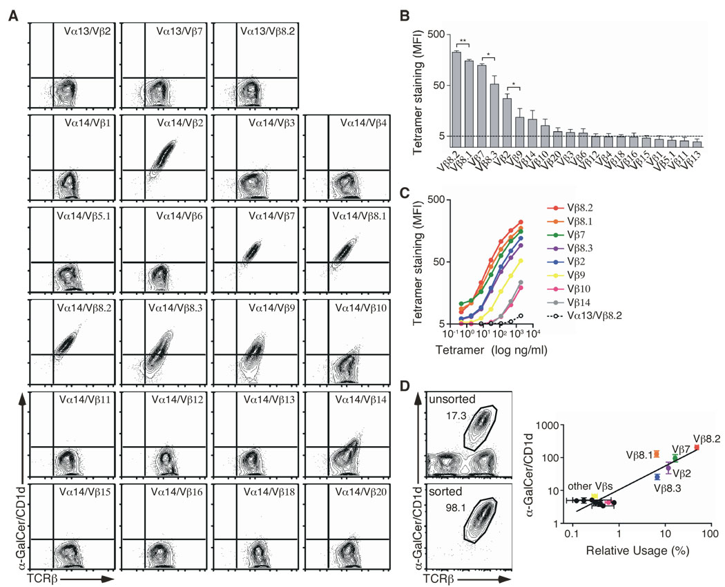
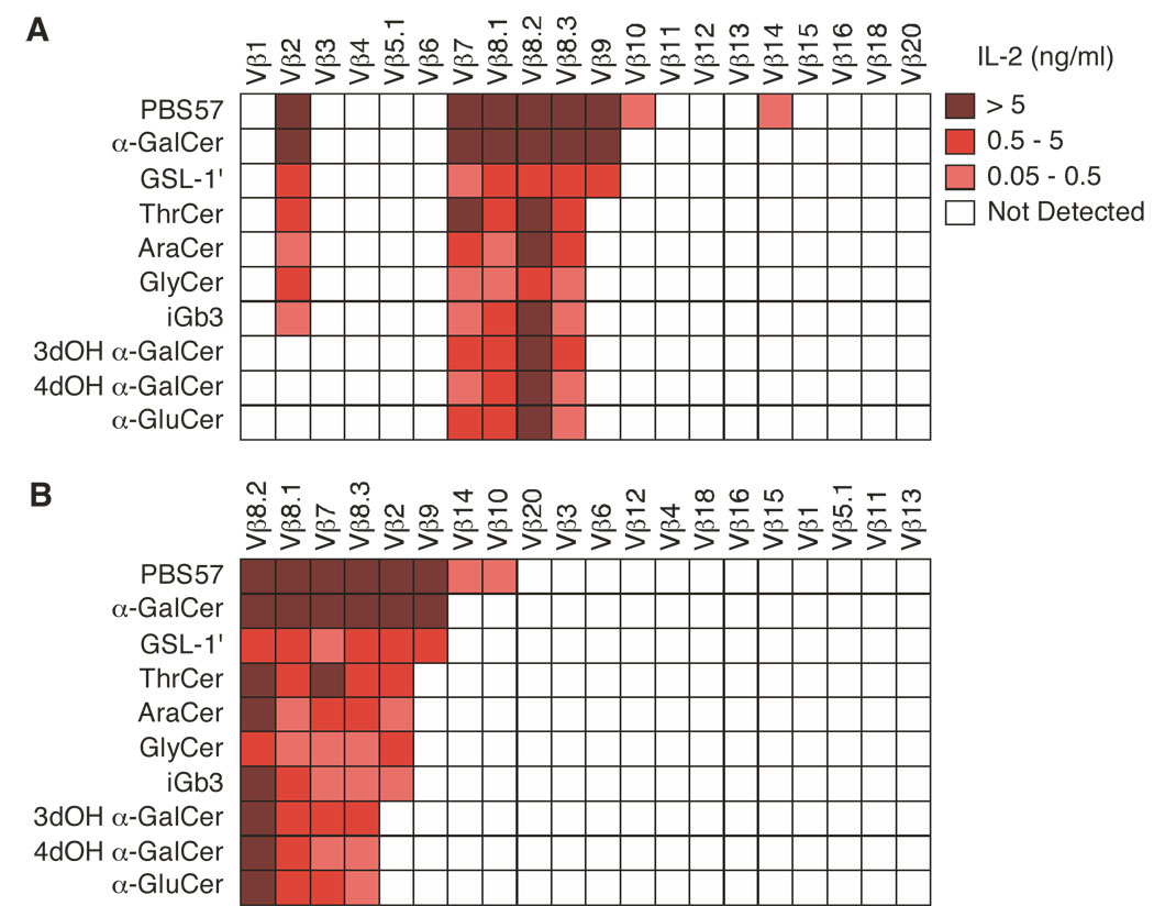
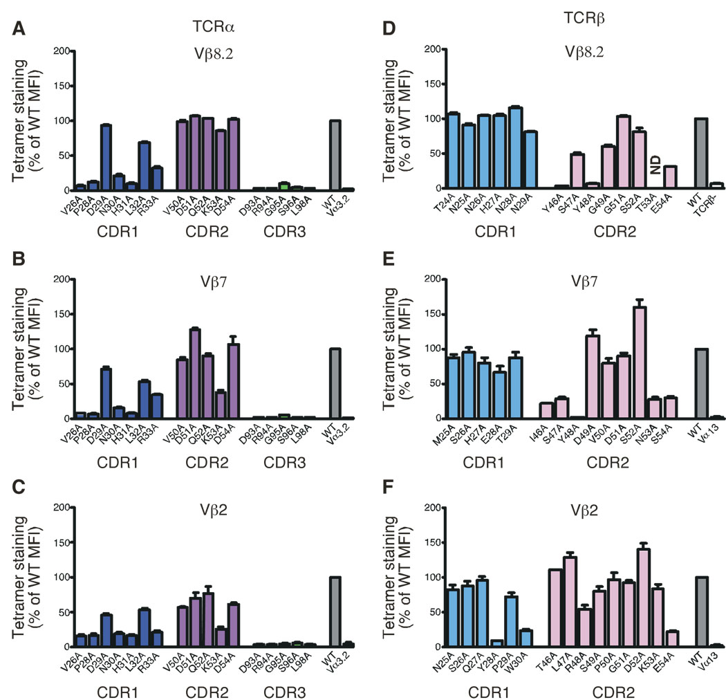
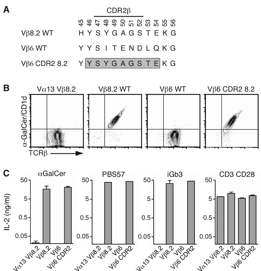
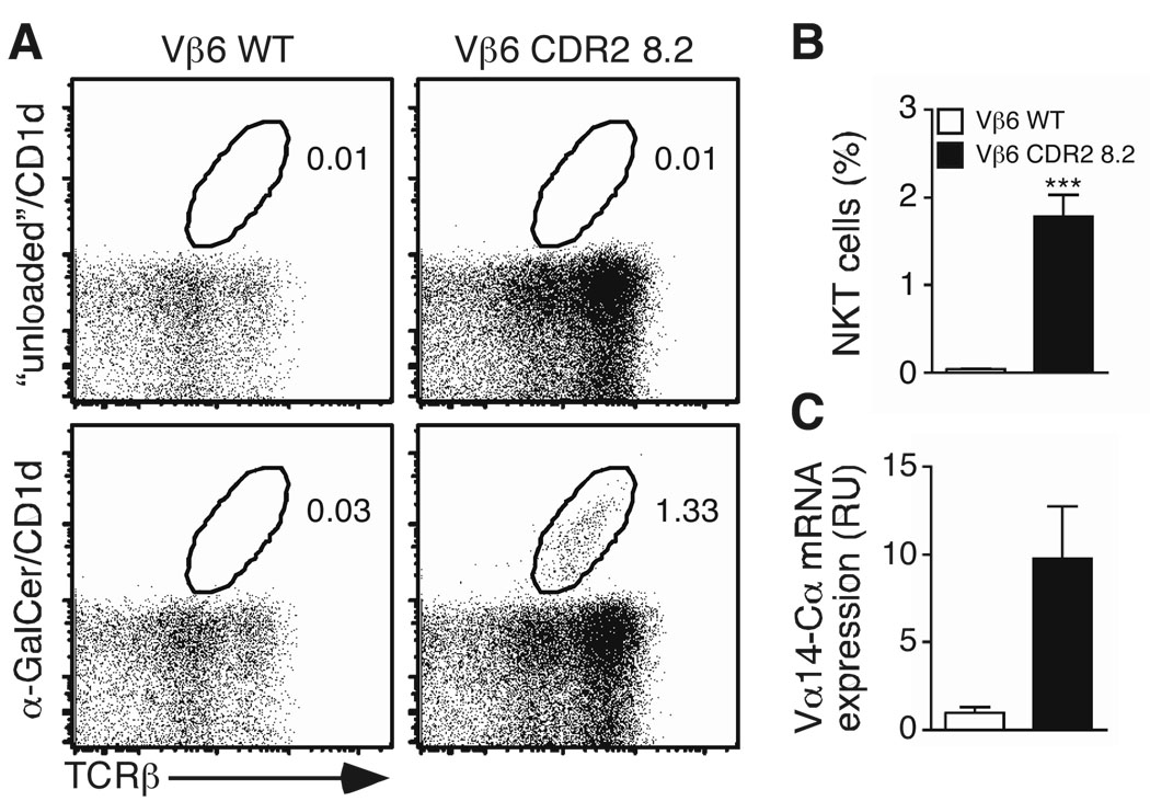
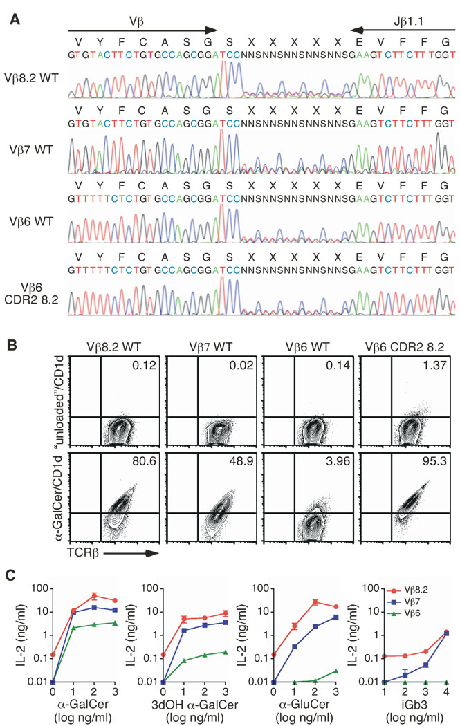
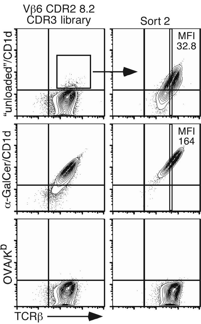
Mouse type I natural killer T cell receptors (iNKT TCRs) use a single V alpha 14-J alpha 18 sequence and V beta s that are almost always V beta 8.2, V beta 7, or V beta 2, although the basis of this differential usage is unclear. We showed that the V beta bias occurred as a consequence of the CDR2 beta loops determining the affinity of the iNKT TCR for CD1d-glycolipids, thus controlling positive selection. Within a conserved iNKT-TCR-CD1d docking framework, these inherent V beta-CD1d affinities are further modulated by the hypervariable CDR3 beta loop, thereby defining a functional interplay between the two iNKT TCR CDR beta loops. These V beta biases revealed a broadly hierarchical response in which V beta 8.2 > V beta 7 > V beta 2 in the recognition of diverse CD1d ligands. This restriction of the iNKT TCR repertoire during thymic selection paradoxically ensures that each peripheral iNKT cell recognizes a similar spectrum of antigens.


Conflict of interest statement

COMPETING INTERESTS STATEMENT

The authors declare no financial conflict of interest.

Figures

Figure 1
Figure 1
Vβ chains confer differential affinity for CD1d. (A) Staining of hybridomas expressing the Vα14i TCRα chain, or Vα13-Jα18 TCRα chain (negative controls), paired with the indicated Vβ chains in the context of a unique CDR3β. (B) The MFI of αGC/CD1d tetramer staining for each hybridoma was determined for a narrow TCR gate. Data represent the mean + s.e.m. of three independent experiments. (C) The indicated hybridomas were stained with increasing concentrations of αGC/CD1d tetramer. The MFI of tetramer staining was determined for a narrow TCR gate. The data represent the mean of two independent experiments. (D) Ex vivo sorted NKT cells were stained with specific anti-Vβ antibodies. Data represent a plot of the relative Vβ usage (x axis) against the αGC/CD1d tetramer MFI of the appropriate Vβ-expressing hybridomas as determined in (B) (y axis). Data represent the mean ± s.e.m. of two independent experiments.
Figure 2
Figure 2
Clustering of Vβ-containing iNKT TCRs according to antigen stimulation. (A, B) Enzyme-linked immunosorbent assay of IL-2 production by hybridomas expressing iNKT TCRs containing Vβ1 to Vβ20 TCRβ chains, stimulated with mCD1d-expressing A20 cells in the presence of PBS57 (1 µg/ml), αGC (1 µg/ml), GSL-1’ (1 µg/ml), ThrCer (0.2 µg/ml), AraCer (0.2 µg/ml), GlyCer (0.2 µg/ml), iGb3 (10 µg/ml), 3dOH αGC (1 µg/ml), 4dOH αGC (1 µg/ml) and α-GluCer (1 µg/ml). (B) Rearrangement of the strength of the response versus the number of hybridomas stimulated. Data represent the mean of two to four independent experiments.
Figure 3
Figure 3
Mutational analysis of Vβ8.2, Vβ7 and Vβ2-containing iNKT TCRs. Staining of hybridomas expressing mutant versions of the Vα14i TCRα chain associated with the wild-type (A) Vβ8.2, (B) Vβ7 or (C) Vβ2 TCRβ chains. Staining of hybridomas expressing alanine substitutions of (D) Vβ8.2, (E) Vβ7 or (F) Vβ2 associated with the wild-type Vα14i TCRα chain. Dark blue, CDR1α; magenta, CDR2α; green, CDR3α, light blue, CDR1β and pink, CDR2β. WT, unsubstituted Vα14i TCRα chain paired with unsubstituted Vβ8.2, Vβ7 or Vβ2 TCRβ chains (wild-type controls). Vα3.2, Vα14i TCRα chain in which the CDR1α region is swapped for the Vα3.2 CDR1α region, and paired with the appropriate TCRβ chains (negative controls for TCRα substitutions). Vα13, Vα13-Jα18 TCRα chain paired with the appropriate TCRβ chains (negative controls for the TCRβ substitutions). ND, not done. The MFI of tetramer staining for each mutant was determined for a narrow TCR gate and normalized to wild-type MFI (set as 100%). Data represent the mean + s.e.m. of three independent experiments. Analysis of the Vβ8.2 mutants, with the exception of Y46A and E54A mutants, has been published previously in (Scott Browne et al., 2007) and is shown for comparison.
Figure 4
Figure 4
CDR2β swapping restores CD1d-glycolipid recognition. (A) The Vβ6 CDR2β was substituted with that of Vβ8.2, from positions 46 to 54 (boxed sequence). (B) Staining of hybridomas expressing the indicated TCRβ chain associated with the Vα14i TCRα chain. (C) Enzyme-linked immunosorbent assay of IL-2 production by hybridomas stimulated with mCD1d-expressing A20 cells in the presence of αGC (1 µg/ml), PBS57 (1 µg/ml) or iGb3 (10 µg/ml), or plate-bound anti-CD3 (5 µg/ml) and anti-CD28 (2 µg/ml) antibodies. Data represent the mean + s.e.m. of three independent experiments.
Figure 5
Figure 5
CDR2β swapping restores iNKT-cell development in vivo. TCRβ-deficient donor bone marrow cells were infected with retroviruses encoding the indicated TCRβ chain and eGFP as a reporter. Cells were injected i.v. into sub-lethally irradiated CD45.1 congenic recipient mice. (A, B) Thymocytes were stained 5 weeks post-reconstitution. Cells were gated on eGFP+ B220- CD8- F4/80 Gr-1- cells and presented plots are representative of two independent experiments (3 mice/group) (A). Percentage of αGC/CD1d tetramer+/TCRβ+ cells in the thymus of TCRβ retrogenic mice (B) Data shown are the mean percentage + s.e.m. of 6 mice/group. Statistical significance (p < 0.001) was determined using unpaired Student’s t test. (C) Thymocytes from TCRβ retrogenic mice were depleted of cells expressing CD8α and CD45.1 and sorted for eGFP, TCRβ and CD4 expression and total RNA was prepared. Amounts of Vα14-Cα transcripts were analyzed by quantitative PCR. Normalization of the samples was relative to the quantity of Cα transcripts. Data are representative of two experiments.
Figure 6
Figure 6
CDR3β modulates iNKT TCR affinity. (A) Sequence of the wild-type Vβ8.2, wild-type Vβ7, wild-type Vβ6 WT and CDR2-modified Vβ6 CDR3β random constructs. (B) Staining of hybridomas expressing the indicated CDR3β random construct associated with the Vα14i TCRα chain. The percentage of αGC/CD1d tetramer+ cells is shown. Plots are gated on live eGFP+ cells and are representative of four independent experiments. (C) Enzyme-linked immunosorbent assay of IL-2 production by hybridomas stimulated with the indicated antigen in the presence of mCD1d-expressing A20 cells. One representative experiment out of two is shown.
Figure 7
Figure 7
Optimal CDR2β and CDR3β composition improves iNKT TCR affinity for CD1d. “Unloaded”/CD1d tetramer+ hybridomas derived from the CDR2-modified Vβ6 chain TCR library were sorted twice and stained with the indicated tetramers. Plots are gated on live eGFP+ cells and are representative of three independent experiments.

References

    1. Adams EJ, Strop P, Shin S, Chien YH, Garcia KC. An autonomous CDR3™ is sufficient for recognition of the nonclassical MHC class I molecules T10 and T22 by ©™ T cells. Nat Immunol. 2008;9:777–784. - PMC - PubMed
    1. Apostolou I, Cumano A, Gachelin G, Kourilsky P. Evidence for two subgroups of CD4−CD8− NKT cells with distinct TCRαβ repertoires and differential distribution in lymphoid tissues. J Immunol. 2000;165:2481–2490. - PubMed
    1. Arden B, Clark SP, Kabelitz D, Mak TW. Mouse T-cell receptor variable gene segment families. Immunogenetics. 1995;42:501–530. - PubMed
    1. Behar SM, Podrebarac TA, Roy CJ, Wang CR, Brenner MB. Diverse TCRs recognize murine CD1. J Immunol. 1999;162:161–167. - PubMed
    1. Bendelac A, Savage PB, Teyton L. The Biology of NKT Cells. Annu Rev Immunol. 2006 - PubMed

Publication types

MeSH terms

Substances