. 2009 Jul 17;31(1):47-59.
doi: 10.1016/j.immuni.2009.04.018.

Differential recognition of CD1d-alpha-galactosyl ceramide by the V beta 8.2 and V beta 7 semi-invariant NKT T cell receptors

Affiliations

Differential recognition of CD1d-alpha-galactosyl ceramide by the V beta 8.2 and V beta 7 semi-invariant NKT T cell receptors

Daniel G Pellicci et al. Immunity. .

Abstract

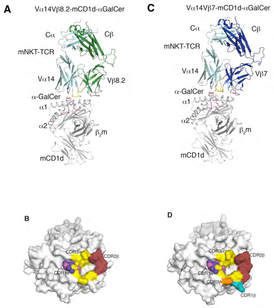
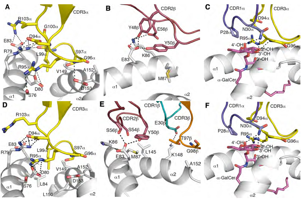
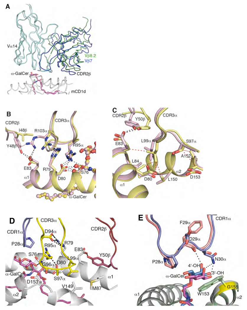
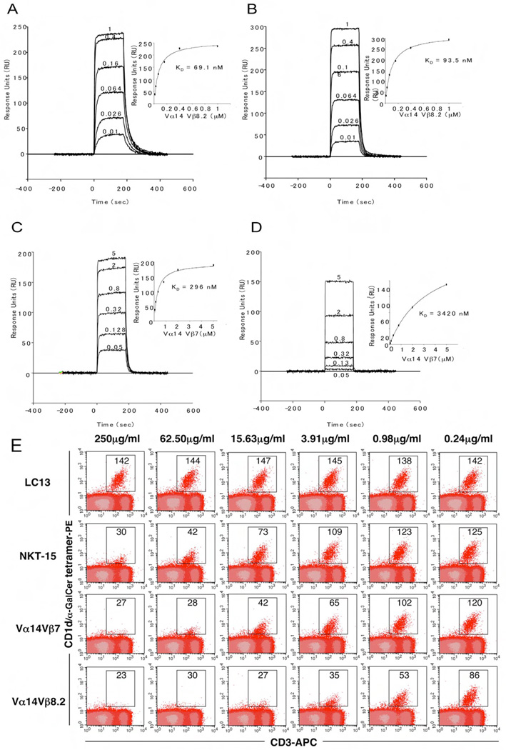
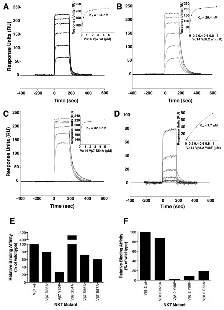
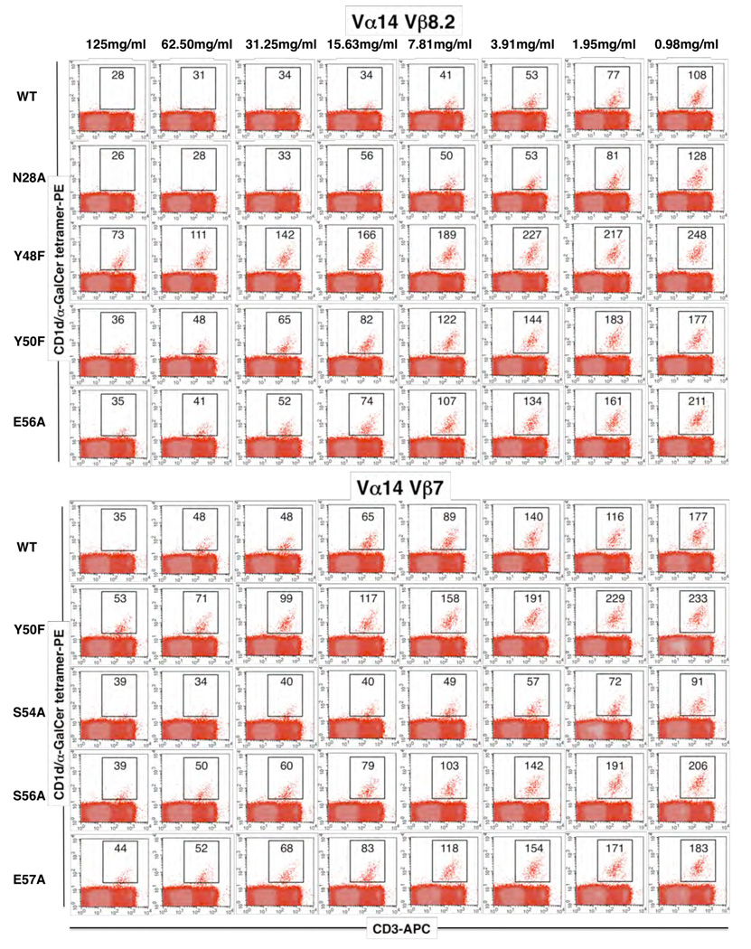
The semi-invariant natural killer T cell receptor (NKT TCR) recognizes CD1d-lipid antigens. Although the TCR alpha chain is typically invariant, the beta chain expression is more diverse, where three V beta chains are commonly expressed in mice. We report the structures of V alpha 14-V beta 8.2 and V alpha 14-V beta 7 NKT TCRs in complex with CD1d-alpha-galactosylceramide (alpha-GalCer) and the 2.5 A structure of the human NKT TCR-CD1d-alpha-GalCer complex. Both V beta 8.2 and V beta 7 NKT TCRs and the human NKT TCR ligated CD1d-alpha-GalCer in a similar manner, highlighting the evolutionarily conserved interaction. However, differences within the V beta domains of the V beta 8.2 and V beta 7 NKT TCR-CD1d complexes resulted in altered TCR beta-CD1d-mediated contacts and modulated recognition mediated by the invariant alpha chain. Mutagenesis studies revealed the differing contributions of V beta 8.2 and V beta 7 residues within the CDR2 beta loop in mediating contacts with CD1d. Collectively we provide a structural basis for the differential NKT TCR V beta usage in NKT cells.


Figures

Figure 1
Figure 1
Structure of mouse NKT TCRs in complex with mouse CD1d-α-GalCer (A) Vα14-Vβ8.2 NKT TCR in complex with mCD1d-α-GalCer. α-GalCer, magenta; mCD1d heterodimer, grey; TCR α-chain, cyan; Vβ8.2 NKT TCR β-chain, green; CDR1α, purple; CDR3α, yellow; CDR1β, teal; CDR2β, ruby; CDR3β, orange; mobile CDR3β region, dashed orange. (B) Footprint of the Vα14-Vβ8.2 NKT TCR on the surface of mouse CD1d-α-GalCer. α-GalCer is shown in spheres. mCD1d, α-GalCer and CDR loops colour coding as in A. (C) Vα14-Vβ7 NKT TCR in complex with mouse CD1d-α-GalCer. Vβ7 NKT TCR β-chain, blue. TCR α-chain, mCD1d, CDR loops and α-GalCer colour coding as in A. (D) Footprint of the Vα14-Vβ7 NKT TCR on the surface of mCD1d-α-GalCer. α-GalCer is shown in spheres. mCD1d, α-GalCer and CDR loops colour coding as in A.
Figure 2
Figure 2
Mouse CD1d and α-GalCer mediated interactions with mouse NKT TCRs CDR3α mediates multiple contacts between mCD1d α-helices and α-GalCer. CDR2β contacts α1-helix of mCD1d. CDR1α interacts solely with α-GalCer galactose head group. CDR1β mediates polar interactions with the α2-helix only in Vβ7 NKT TCR-mCD1d-α-GalCer. (A) Vβ8.2 NKT TCR CDR3α contacts with mCD1d. (B) Vβ8.2 NKT TCR CDR2β contacts with mCD1d. (C) Vβ8.2 NKT TCR CDR1α and CDR3α contacts with α-GalCer. (D) Vβ7 NKT TCR CDR3α contacts with mCD1d. (E) Vβ7 NKT TCR CDR1β, CDR2β and CDR3β contacts with mCD1d. (F) Vβ7 NKT TCR CDR1α and CDR3α contacts with α-GalCer. CDR1α, purple; CDR3α, yellow; CDR1β, teal; CDR2β, ruby; CDR3β, orange; α-GalCer, magenta; mCD1d, grey. H-bond or salt-bridge interactions are shown in black dashed lines.
Figure 3
Figure 3
Comparison of Vα14-Vβ8.2, Vα14-Vβ7 and Vα24-Vβ11 NKT TCR-mCD1d-α-GalCer complexes (A) Superposition of Vα14-Vβ8.2 NKT TCR-mCD1d-α-GalCer and Vα14-Vβ7 NKT TCR-mCD1d-α-GalCer. Differences in the relative juxta-positioning of the Vβ8.2-Vβ7 and Vα14 domains. TCR α-chain, cyan; Vβ8.2 NKT TCR β-chain, green; Vβ7 NKT TCR β-chain, blue; α-GalCer, magenta; mCD1d, grey (B) Differences in the sequence of CDR2β in Vβ8.2 and Vβ7 NKT TCR affected the position of Arg 103α in the CDR3α loop and subsequently altered positions and contacts of Arg 79, Asp 80, Ser 76 and Arg 95α. Vα14-Vβ8.2 NKT TCR-mCD1d-α-GalCer, pink; Vα14-Vβ7 NKT TCR-mCD1d-α-GalCer, yellow. α-GalCer is shown in ball and stick. H-bond or salt-bridge interactions are shown in black dashed lines and vdw interactions are shown in red dashed lines. (C) Altered position of Tyr 50β in Vβ7 NKT TCR affected contacts made by Ser 97α and Leu 99α at the tip of CDR3α with mCD1d. Colour coding as in B. H-bonds are shown in black dashed lines and vdw interactions are shown in red dashed lines. (D) Conserved interactions mediated by CDR1α, CDR3α and CDR2β loops of the human and mouse NKT TCRs on the surface of CD1d and α-GalCer. CDR1α, purple; CDR2β, ruby; CDR3α, yellow; α-GalCer, magenta; CD1d, grey. The numbering shown on CD1d is according to the mouse CD1d. H-bonds or salt-bridge interactions are shown in black dashed lines. (E) The shift in the position of the galactose head group of α-GalCer between mouse and the human NKT TCR-CD1d-α-GalCer structures is due to the presence of a bulky tryptophan side chain in human CD1d (Trp 153) in contrast to glycine (Gly 155, shown in yellow) in mouse CD1d. Human CDR1α, salmon; mouse CDR1α, purple; α-GalCer in human, marine; α-GalCer in mouse, magenta; hCD1d, pale green; mCD1d, grey.
Figure 4
Figure 4
Differential binding affinities of NKT TCRs to CD1d-α-GalCer. Vα14Jα18-Vβ8.2 (A and B) and Vα14Jα18-Vβ7 (C and D) NKT TCR were injected over streptavidin immobilised mouse (A and C) and human (B and D) CD1d-α–GalCer and simultaneously over a control cell coated with unloaded CD1d. Sensograms show the binding (response units, RU) of increasing concentrations of TCR (0.01 to 1µM for Vα14Jα18-Vβ8.2 and 0.05 to 5 µM for Vα14Jα18-Vβ7) to mouse and human CD1d-α–GalCer following baseline subtraction. Insets show saturation plots demonstrating equilibrium binding of NKT TCR to immobilised CD1d-α–GalCer. The equilibrium dissociation constants (KD) derived by equilibrium analysis were equivalent to those derived by kinetic analysis. E) CD1d-α–GalCer tetramer inhibition. Recombinant soluble NKT TCRs were examined for their ability to block binding of mCD1d/αGC tetramers to mouse NKT cells. PE labelled CD1d-α–GalCer tetramers were pre-incubated with titrating amounts of soluble NKT TCRs or an irrelevant TCR control, LC13, before staining of mouse thymocytes. Cells were analysed by flow cytometry showing mCD1d-α–GalCer tetramer-PE on the vertical axis and anti-CD3 APC on the horizontal axis. CD3+ mCD1d-α–GalCer tetramer+ thymic NKT cells are indicated within the square with the MFI (mean fluorescence intensity) indicated. All measurements were taken in duplicate.
Figure 5
Figure 5
Binding of mutant NKT TCRs to mouse CD1d-α-GalCer as assessed by surface plasmon resonance. Wild type Vβ7 NKT TCR (A) and Vβ8.2 NKT TCR (B) and mutant Vβ7 NKT TCR S54A (C) and mutant Vβ8.2 (Y48F) (D) NKT TCR were injected over streptavidin immobilised mouse CD1d-α-GalCer and over a control cell containing unloaded CD1d. Sensorgrams show the binding (response units, RU) of decreasing concentrations of TCR (5, 2, 0.8, 0.32, 0.13 and 0.05 for Vα14Jα18-Vβ7 TCRs and 1, 0.4, 0.16, 0.064, 0.026 and 0.01µM for Vα14Jα18-Vβ8.2 TCRs) to mouse CD1d-α-GalCer following subtraction of the control flow cell. Insets show saturation plots demonstrating equilibrium binding of NKT TCR to immobilised CD1d-α-GalCer. (E and F) Binding of mutant NKT TCR to mouse CD1d α-GalCer. Site directed mutants of individual Vβ7 or Vβ8.2 residues were refolded with the invariant α-chain. The data is presented as a percentage binding of wild-type NKT TCRTCR.
Figure 6
Figure 6
Binding of mutant NKT TCRs as assessed by CD1d-α-GalCer tetramer inhibition. Recombinant soluble NKT TCRs, and mutants thereof, were examined for their ability to block binding of mCD1d-α-GalCer tetramers to mouse NKT cells. PE labelled CD1d-α-GalCer tetramers were pre-incubated with titrating amounts of soluble wild type and mutant NKT TCRs before staining of mouse thymocytes. Cells were analysed by flow cytometry showing mCD1d-α–GalCer tetramer-PE on the vertical axis and anti-CD3 APC on the horizontal axis. CD3+ mCD1d-α–GalCer tetramer+ thymic NKT cells are indicated within the square with the MFI indicated.

Similar articles

Cited by

References

    1. Bendelac A, Savage PB, Teyton L. The Biology of NKT Cells. Annu Rev Immunol. 2007;25:297–336. - PubMed
    1. Benlagha K, Weiss A, Beavis A, Teyton L, Bendelac A. In vivo identification of glycolipid antigen-specific T cells using fluorescent CD1d tetramers. Journal of Experimental Medicine. 2000;191:1895–1903. - PMC - PubMed
    1. Borg NA, Wun KS, Kjer-Nielsen L, Wilce MC, Pellicci DG, Koh R, Besra GS, Bharadwaj M, Godfrey DI, McCluskey J, Rossjohn J. CD1d-lipid-antigen recognition by the semi-invariant NKT T-cell receptor. Nature. 2007;448:44–49. - PubMed
    1. Brigl M, van den Elzen P, Chen X, Meyers JH, Wu D, Wong CH, Reddington F, Illarianov PA, Besra GS, Brenner MB, Gumperz JE. Conserved and Heterogeneous Lipid Antigen Specificities of CD1d-Restricted NKT Cell Receptors. J Immunol. 2006;176:3625–3634. - PubMed
    1. Brossay L, Chioda M, Burdin N, Koezuka Y, Casorati G, Dellabona P, Kronenberg M. Cd1d-Mediated Recognition of an Alpha-Galactosylceramide by Natural Killer T Cells Is Highly Conserved through Mammalian Evolution. Journal of Experimental Medicine. 1998;188:1521–1528. - PMC - PubMed

Publication types

MeSH terms