. 2009 Sep 11;284(37):24881-90.
doi: 10.1074/jbc.M109.014365. Epub 2009 Jul 10.

Cell polarity factor Par3 binds SPTLC1 and modulates monocyte serine palmitoyltransferase activity and chemotaxis

Affiliations

Cell polarity factor Par3 binds SPTLC1 and modulates monocyte serine palmitoyltransferase activity and chemotaxis

Norimasa Tamehiro et al. J Biol Chem. .

Abstract

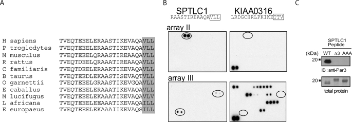
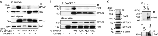
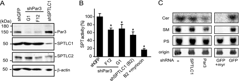
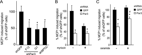
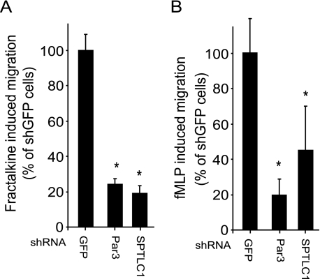
Elevated sphingolipids have been associated with increased cardiovascular disease. Conversely, atherosclerosis is reduced in mice by blocking de novo synthesis of sphingolipids catalyzed by serine palmitoyltransferase (SPT). The SPT enzyme is composed of the SPTLC1 and -2 subunits, and here we describe a novel protein-protein interaction between SPTLC1 and the PDZ protein Par3 (partitioning defective protein 3). Mammalian SPTLC1 orthologs have a highly conserved C terminus that conforms to a type II PDZ protein interaction motif, and by screening PDZ domain protein arrays with an SPTLC1 C-terminal peptide, we found it bound the third PDZ domain of Par3. Overlay and immunoprecipitation assays confirmed this interaction and indicate Par3 is able to associate with the SPTLC1/2 holoenzyme by binding the C-terminal SPTLC1 PDZ motif. The physiologic existence of the SPTLC1/2-Par3 complex was detected in mouse liver and macrophages, and short interfering RNA inhibition of Par3 in human THP-1 monocytes significantly reduced SPT activity and de novo ceramide synthesis by nearly 40%. Given monocyte recruitment into inflamed vessels is thought to promote atherosclerosis, and because Par3 and sphingolipids have been associated with polarized cell migration, we tested whether the ability of THP-1 monocytes to migrate toward MCP-1 (monocyte chemoattractant protein 1) depended upon Par3 and SPTLC1 expression. Knockdown of Par3 significantly reduced MCP1-induced chemotaxis of THP-1 monocytes, as did knockdown of SPTLC1, and this Par3 effect depended upon SPT activity and was blunted by ceramide treatment. In conclusion, protein arrays were used to identify a novel SPTLC1-Par3 interaction that associates with increased monocyte serine palmitoyltransferase activity and chemotaxis toward inflammatory signals.


Figures

FIGURE 1.
FIGURE 1.
SPTLC1 contains a conserved C-terminal PDZ motif that interacts with the third PDZ domain of Par3. A, alignment of the C-terminal 25 amino acids of SPTLC1 from the indicated species is shown, and the final three conserved hydrophobic residues that conform to a class II PDZ motif (XΦ−2X1Φ0-COOH) are boxed. B, SPTLC1 C terminus is able to bind to the third PDZ domain of Par3 as identified by the binding of a biotinylated 20-mer C-terminal SPTLC1 peptide to protein arrays that are spotted in duplicate with 123 PDZ domains from 73 proteins. Shown in the left panels are the two arrays of four containing PDZ domains that bound the SPTLC1 peptide (signal intensity FRMPD4 > Par3 > PTPN13). That binding of the SPTLC1 peptide to these PDZ domains was driven by a type II interaction was indicated by the binding pattern of a control biotinylated peptide (KIAA0316), which conforms to a class I PDZ motif (X(S/T)−2X10-COOH) and interacts with a distinct set of PDZ domains (right panels). C, binding of full-length Par3 (150-kDa isoform) to the SPTLC1 C terminus requires the final three residues that comprise the SPTLC1 type II PDZ motif as determined by overlay assays. Purified bacterial polypeptides encoding the wild type SPTLC1 C terminus or the C terminus with the PDZ motif deleted (Δ3) or substituted with alanines (AAA) were separated by SDS-PAGE and transferred to nitrocellulose. Incubation of the membrane with a lysate containing HA-tagged Par3 expressed in 293-EBNA-T cells only bound WT SPTLC1 as determined by immunoblotting with an anti-HA antibody. Gels run in parallel and Coomassie-stained for total SPTLC1 protein indicate the failure of Par3 to bind the mutant SPTLC1 polypeptides was not due to unequal loading in these assays.
FIGURE 2.
FIGURE 2.
Par3 binding of SPTLC1 requires the SPTLC1 type II PDZ motif and occurs at physiologic expression levels in the mouse liver. A, immunoprecipitation (IP) of HA-Par3 out of 293-EBNA-T cells expressing FLAG (FL)-tagged SPTLC1 or the indicated FLAG-SPTLC1 mutants showing the SPTLC1 C-terminal PDZ motif is required for the binding of SPTLC1 and Par3 in a cellular context. The top two panels show immunoblots (IB) of the precipitated SPTLC1 and Par3, and the lower two panels show the amount of total SPTLC1 and Par3 expressed in the transiently transfected 293HEK cells. B, converse immunoprecipitations of the FLAG-SPTLC1 proteins confirm the Par3 interaction with SPTLC1 requires the SPTLC1 C-terminal PDZ motif. C, Par3 150-kDa isoform is expressed in the mouse liver along with the SPTLC1 and SPTLC2 subunits as determined by immunoblots (left panels) and interacts with SPTLC1 and SPTLC2 as determined by immunoprecipitation of the liver lysates with normal IgG or antibodies against SPTLC1 (top right panel) or SPTLC2 (lower right panel).
FIGURE 3.
FIGURE 3.
Par3 interacts with the SPTLC1/2 holoenyzme through an interaction with the SPTLC1 PDZ motif. Myc-tagged SPTLC2 was co-transfected into 293-EBNA-T cells alone or in the presence of HA-Par3 and the indicated FLAG-SPTLC1 constructs. SPTLC2 was immunoprecipitated (IP) with an anti-Myc antibody, and the amount of Par3, SPTLC1, and SPTLC2 that precipitated was determined by immunoblotting (IB) (left panels), and immunoblotting the input protein lysates shows the total amount of Par3, SPTLC1, and SPTLC2 that was expressed in the cells (right panels).
FIGURE 4.
FIGURE 4.
shRNA suppression of Par3 in human THP-1 monocytes inhibits serine palmitoyltransferase activity. A, THP-1 monocytes express Par3, SPTLC1, and SPTLC2 as determined by immunoblotting, and lentivirus-mediated expression of shRNAs targeting Par3 (G1 and F12) or SPTLC1 specifically repress Par3 and SPTLC1 protein expression, respectively. B, lentivirus delivered shRNAs targeting Par3, and SPTLC1 inhibits SPT activity by ∼40% in THP-1 monocytes as determined by measuring the condensation of [14C]serine and palmitoyl-CoA catalyzed by microsomal cell membrane lysates. Pretreatment of the SPTLC1-targeted lysate with the SPT inhibitor myriocin further inhibited activity to 90% (B2 + myriocin, n = 3, ±S.D., *, p < 0.05). C, de novo synthesis of ceramide (Cer) and sphingomyelin (SM) is reduced in cells expressing shRNAs targeting Par3 or SPTLC1 expression as compared with uninfected control cells, or cells expressing a lentivirus-delivered shRNA targeting GFP. As with the Par3- and SPTLC1-targeted cells, myriocin treatment of the control GFP-targeted cells reduced incorporation of [14C]serine into ceramide (Cer) and sphingomyelin (SM) but not phosphatidylserine (PS). Cells were incubated with [14C]serine (5 μCi) for 24 h at 37 °C, and cellular lipids were isolated by chloroform/methanol extraction and separated on TLC plates along with purified standards for ceramide, sphingomyelin, and phosphatidylserine. Labeled lipids remaining at the plate origin was similar demonstrating equal loading of the samples (results representative of two or more independent experiments, and quantification of labeled ceramide and sphingomyelin is shown in supplemental Fig. 5B).
FIGURE 5.
FIGURE 5.
shRNA suppression of Par3 and SPTLC1 inhibits polarized migration of THP-1 cells in response to monocyte chemoattractant protein 1. A, THP-1 monocytes expressing shRNAs against Par3 or SPTLC1 have an ∼50% reduction in their ability to migrate toward a gradient of MCP-1 (10 nm) compared with control cells expressing an shRNA targeting GFP as determined by Boyden chamber assays. B, myriocin pretreatment of control cells expressing the GFP shRNA reduces their MCP-1-induced migration by 44% but does affect the residual MCP-1-induced migration of THP-1 monocytes with suppressed Par3 expression. C, ceramide pretreatment of Par3-suppressed THP-1 monocytes significantly improves their MCP-1-induced migration defect (n = 3, ± S.D., *, p < 0.05 versus shGFP untreated; #, p < 0.05 versus shPar3 untreated; results representative of two or more independent experiments).
FIGURE 6.
FIGURE 6.
Loss of Par3 or SPTLC1 expression inhibits THP-1 chemotaxis induced by structurally diverse chemokines including Fractalkine and the fMLP bacterially formylated tripeptide. A, Fractalkine (10 nm)-induced migration of THP-1 cells expressing shRNAs against Par3 or SPTLC1 is reduced by ∼80% compared with control cells expressing an shRNA targeting GFP. B, fMLP (10 nm)-induced migration of THP-1 cells expressing shRNAs against SPTLC1 or Par3 is reduced ∼60 and 80%, respectively, as compared with the control GFP shRNA cells (n = 3, ±S.D., *, p < 0.05; results are representative of two or more independent experiments).

References

    1. Rivera J., Proia R. L., Olivera A. (2008) Nat. Rev. Immunol. 8, 753–763 - PMC - PubMed
    1. van Meer G., Voelker D. R., Feigenson G. W. (2008) Nat. Rev. Mol. Cell Biol. 9, 112–124 - PMC - PubMed
    1. Wymann M. P., Schneiter R. (2008) Nat. Rev. Mol. Cell Biol. 9, 162–176 - PubMed
    1. Zuo Y., Zhuang D. Z., Han R., Isaac G., Tobin J. J., McKee M., Welti R., Brissette J. L., Fitzgerald M. L., Freeman M. W. (2008) J. Biol. Chem. 283, 36624–36635 - PMC - PubMed
    1. Hanada K. (2003) Biochim. Biophys. Acta 1632, 16–30 - PubMed

Publication types