. 2009 Jul 28;106(30):12465-70.
doi: 10.1073/pnas.0905931106. Epub 2009 Jul 10.

ETS family transcription factors collaborate with alternative signaling pathways to induce carcinoma from adult murine prostate cells

Affiliations

ETS family transcription factors collaborate with alternative signaling pathways to induce carcinoma from adult murine prostate cells

Yang Zong et al. Proc Natl Acad Sci U S A. .

Abstract

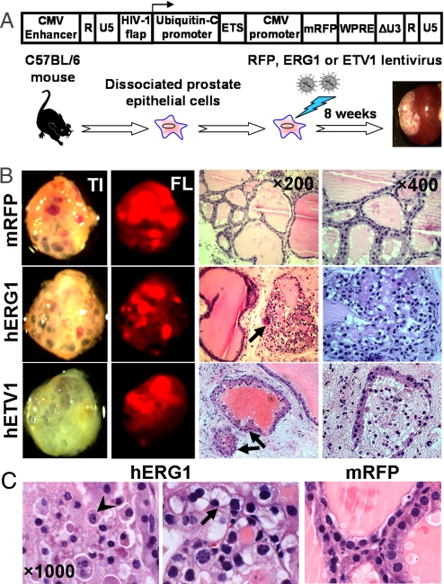
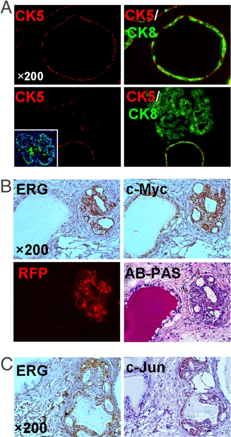
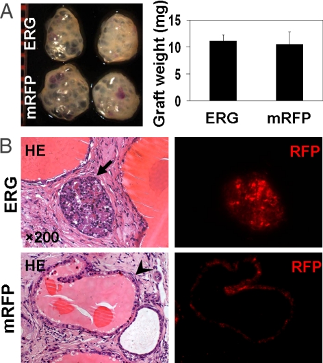
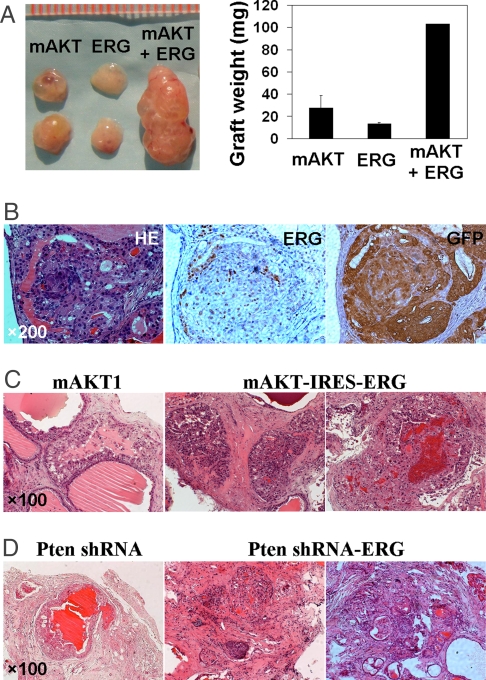
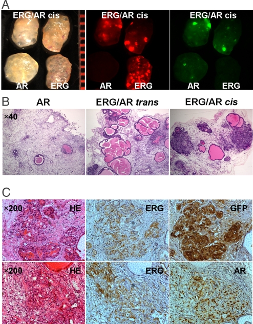
Chromosomal rearrangements involving erythroblast transformation specific (ETS) family transcription factors were recently defined as the most common genetic alterations in human prostate cancer. Despite their prevalence, it is unclear what quantitative role they play in either initiation or progression of the disease. Using a lentiviral transduction and dissociated cell prostate regeneration approach, we find that acutely increased expression of ETS proteins in adult murine prostate epithelial cells is sufficient to induce the formation of epithelial hyperplasia and focal prostatic intraepithelial neoplasia (PIN) lesions, but not progression to carcinoma. However, combined expression of ERG with additional genetic alternations associated with human prostate cancer can lead to aggressive disease. Although ERG overexpression does not cooperate with loss of the tumor suppressor p53, it does collaborate with alterations in PI3K signaling, such as Pten knockdown or AKT up-regulation, to produce a well-differentiated adenocarcinoma. Most striking is our finding that overexpression of androgen receptor (AR) does not give rise to any hyperplastic lesions, but when combined with high levels of ERG, it promotes the development of a more poorly differentiated, invasive adenocarcinoma. These findings suggest that in human prostate cancer, the most potent function of ETS gene fusions may be to synergize with alternative genetic events and provide different pathways for carcinoma production and invasive behavior. Our results provide direct evidence for selective cooperating events in ERG-induced prostate tumorigenesis and offer a rational basis for combined therapeutic interventions against multiple oncogenic pathways in prostate cancer.


Conflict of interest statement

The authors declare no conflict of interest.

Figures

Fig. 1.
Fig. 1.
Epithelial hyperplasia and focal PIN lesions in prostate grafts regenerated from ETS-transduced adult murine prostate cells. (A) Diagram of the ETS lentiviral vector used in the in vivo prostate regeneration assay. Dissociated prostate cells from C57BL/6 mice at 8–12 weeks of age were transduced with the indicated lentivirus and subsequently engrafted into the subrenal capsules of SCID mice and regenerated in vivo for 8 weeks. (B) Representative transilluminated (TI), fluorescent (FL) photographs, and histological sections (H&E staining) of regenerated tissues derived from ETS-transduced prostate cells. ETS-transduced prostate tubules are indicated by arrows. (C) Cellular and nuclear atypia and signet ring cell-like appearance of the epithelial cells in ERG-transduced prostate glands (arrowhead, prominent nucleoli; arrow, intracytoplasmic vacuoles and nuclear displacement). mRFP-tranduced glands are shown as the control.
Fig. 2.
Fig. 2.
ERG overexpression resulted in up-regulation of c-Myc and c-Jun proteins and a skewed cell lineage composition in regenerated prostate tubules. (A) IF analyses of ERG-transduced prostate graft sections (Lower) showing the nuclear staining of ERG in prostate epithelial cells (Inset) and loss of CK5-positive cells in the corresponding tubules compared with the adjunct normal tubules and mRFP-transduced glands (Upper). (B) IHC analysis with anti-ERG and c-Myc antibodies identifies increased levels of c-Myc expression in ERG-positive prostate tubules (Upper). Alcian blue-periodic acid Schiff (AB-PAS) staining and fluorescent (RFP) image of tissue sections containing the same tubules are shown (Lower). (C) IHC staining of regenerated prostate tissues with anti-ERG and c-Jun antibodies reveals the close correlation of ERG overexpression with up-regulation of c-Jun protein.
Fig. 3.
Fig. 3.
The absence of synergy between ERG overexpression and p53 deletion for PCa progression. (A) Macroscopic photo and weight of regenerated tissues derived from ERG or mRFP-transduced p53-null murine prostate cells. (B) Representative histological sections of the graft tissues regenerated from p53-null prostate cells, showing that combined ERG overexpression and loss of p53 in murine prostate cells only resulted in PIN lesions, whereas mRFP-transduced p53−/− prostate epithelial cells gave rise to the normal prostate glands. Arrows indicated the lentiviral-transduced prostate tubules. The fluorescent (RFP) images of tissue sections containing the same tubules are also shown.
Fig. 4.
Fig. 4.
Invasive prostate adenocarcinoma induced by ERG overexpression and aberrant PI3K pathway. (A) Photographic overview and weight of regenerated tissues derived from wild-type prostate cells transduced with the indicated lentiviruses. (B) H&E staining and IHC analyses with antibodies against ERG and GFP show prostate adenocarcinoma developed in ERG/mAKT1 coinfected prostate grafts. (C) H&E staining of tissue sections showing invasive adenocarcinomas induced by ERG overexpression and activated AKT kinase using a bicistronic vector. PIN lesions induced by mAKT1 alone are shown as the control. (D) H&E staining of regenerated tissue sections reveals that the combination of ERG overexpression and Pten knockdown resulted in prostate adenocarcinoma, as compared with PIN lesions induced by Pten knockdown.
Fig. 5.
Fig. 5.
The synergistic effects between ERG overexpression and enhanced AR signaling for PCa progression. (A) Macroscopic transilluminated and fluorescent photos showing the more solid texture in ERG/AR coinfected prostate grafts, compared with the transparent appearance of ERG-transduced grafts. (B) H&E staining of regenerated tissue sections reveals that combined up-regulation of ERG and AR led to the formation of invasive adenocarcinoma in ERG/AR coinfected (ERG/AR cis) prostate grafts, but not in the separately infected/pooled (ERG/AR trans) prostate grafts. Overexpression of AR alone caused an evident decrease in the number of regenerated prostate tubules. (C) H&E staining and IHC analyses with antibodies against ERG, GFP, and AR of regenerated tissue sections showing invasive prostate adenocarcinoma developed in regenerated tissues derived from ERG/AR coinfected prostate cells.

References

    1. Vogelstein B, Kinzler KW. The multistep nature of cancer. Trends Genet. 1993;9:138–141. - PubMed
    1. Vogelstein B, Kinzler KW. Cancer genes and the pathways they control. Nat Med. 2004;10:789–799. - PubMed
    1. Abate-Shen C, Shen MM. Molecular genetics of prostate cancer. Genes Dev. 2000;14:2410–2434. - PubMed
    1. Look AT. Oncogenic transcription factors in the human acute leukemias. Science. 1997;278:1059–1064. - PubMed
    1. Kumar-Sinha C, Tomlins SA, Chinnaiyan AM. Evidence of recurrent gene fusions in common epithelial tumors. Trends Mol Med. 2006;12:529–536. - PubMed

Publication types

MeSH terms