. 2009 Jul 28;106(30):12524-9.
doi: 10.1073/pnas.0903328106. Epub 2009 Jul 10.

Glial ephrin-A3 regulates hippocampal dendritic spine morphology and glutamate transport

Affiliations

Glial ephrin-A3 regulates hippocampal dendritic spine morphology and glutamate transport

Maria A Carmona et al. Proc Natl Acad Sci U S A. .

Abstract

Increasing evidence indicates the importance of neuron-glia communication for synaptic function, but the mechanisms involved are not fully understood. We reported that the EphA4 receptor tyrosine kinase is in dendritic spines of pyramidal neurons of the adult hippocampus and regulates spine morphology. We now show that the ephrin-A3 ligand, which is located in the perisynaptic processes of astrocytes, is essential for maintaining EphA4 activation and normal spine morphology in vivo. Ephrin-A3-knockout mice have spine irregularities similar to those observed in EphA4-knockout mice. Remarkably, loss of ephrin-A3 or EphA4 increases the expression of glial glutamate transporters. Consistent with this, glutamate transport is elevated in ephrin-A3-null hippocampal slices whereas Eph-dependent stimulation of ephrin-A3 signaling inhibits glutamate transport. Furthermore, some forms of hippocampus-dependent learning are impaired in the ephrin-A3-knockout mice. Our results suggest that the interaction between neuronal EphA4 and glial ephrin-A3 bidirectionally controls synapse morphology and glial glutamate transport, ultimately regulating hippocampal function.


Conflict of interest statement

The authors declare no conflict of interest.

Figures

Fig. 1.
Fig. 1.
Abnormal in vivo dendritic spine morphology in ephrin-A3-knockout mice. (A) Three-dimensional reconstruction of diolistically labeled dendritic spines from CA1 pyramidal neurons of 7-week-old wild-type (+/+) and ephrin-A3-knockout (−/−) mice. Arrows point to examples of mushroom (m), stubby (s), or thin (t) spines. (B–E) Quantification of dendritic spine parameters. The ephrin-A3 knockout mice have similar spine density and width compared with control (+/+ and +/−) mice, but their spines are significantly longer and have longer spine necks (***, P < 0.001, KS and Student's t test). (F) Ephrin-A3-knockout mice have more mushroom-shaped spines and fewer stubby spines than control mice (***, P < 0.001, one-way ANOVA with Bonferroni's post hoc comparisons). Error bars, SEM. (Scale bar, 1 μm.)
Fig. 2.
Fig. 2.
Ephrin-A3 promotes EphA4 phosphorylation in the adult hippocampus. (A) Immunoprecipitation and immunoblot analysis indicates lower EphA4 phosphorylation in the 6- to 8-week-old ephrin-A3-null hippocampus (−/−) compared with wild type (+/+). (B) Quantification of 3 experiments shows that EphA4 phosphorylation in ephrin-A3 knockout mice is ≈50% that in wild-type mice (*, P < 0.05, Student's t test). Error bar, SEM. (C) The expression levels of B-type ephrins, analyzed by immunoblotting after immunoprecipitation with a pan-ephrin-B antibody, are similar in wild-type and ephrin-A3-null hippocampus.
Fig. 3.
Fig. 3.
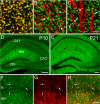
Ephrin-A3 colocalizes with GLAST on astrocytic processes in the adult hippocampus. (A–C) Immunofluorescence confocal images of hippocampal sections from adult mice double-labeled for ephrin-A3 (green) and GLAST (red, A), VGLUT-1 (red, B), and PSD-95 (red, C). Arrows in B and C show examples where ephrin-A3 labeling surrounds presynaptic (B) and postsynaptic (C) glutamatergic terminals. Extensive colocalization with GLAST in A indicates that ephrin-A3 is concentrated in perisynaptic astrocytic processes. (D and E) Overview of ephrin-A3 expression in the hippocampus of P10 and P21 mice. (F–H) Double labeling for ephrin-A3 (F) and GFAP (G) in P10 hippocampus shows extensive colocalization in the merged image (H) (arrows). DG, dentate gyrus; slm, stratum lacunosum moleculare; sr, stratum radiatum. [Scale bars, 3 μm (C); 50 μm (D and E); and 20 μm (H).]
Fig. 4.
Fig. 4.
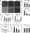
Enhanced levels of glial glutamate transporters in ephrin-A3 and EphA4-knockout mice. (A) Single-plane confocal images showing immunofluorescence labeling for GLT-1, GLAST, and EAAC1 in the CA1 stratum radiatum of the wild-type and ephrin-A3-null hippocampus. (Scale bar, 8 μm.) (B) Quantification of average pixel intensities reveal higher immunoreactivities for GLT-1 and GLAST but not EEAC1 in the ephrin-A3-null hippocampus. (C) The percentage of bright pixels (corresponding to the 5% brightest pixels in the wild-type sections and the pixels as bright or brighter in the knockout sections) indicates higher labeling intensity for GLT-1- and GLAST-positive punctae in the ephrin-A3-knockout mice. (D) Quantification of average pixel intensities in the EphA4-null hippocampus relative to wild type. Average pixel intensities are significantly higher only for GLT-1 in the EphA4-knockout mice compared with wild type. (E) The percentage of bright pixels in the EphA4-knockout hippocampus is higher than wild type for GLT-1, GLAST, and EAAC1, but not ephrin-A3. (F) Immunoblots comparing expression of the indicated proteins in wild-type and ephrin-A3-null hippocampus. (G) Quantification of immunoblots from 4 pairs of wild-type and ephrin-A3-knockout mice by densitometry indicates higher expression of glial transporters in the knockout hippocampus whereas expression of the neuronal glutamate transporter EAAC1 is similar. Values were normalized to GAPDH levels and expressed as a percentage of the levels in the wild-type hippocampus. (H) mRNA levels are similar in the wild-type and ephrin-A3-null hippocampus, as determined by real-time RT-PCR analysis. Values were normalized to GAPDH transcript levels and expressed as a percentage of the levels in wild-type hippocampus. Error bars, SEM; *, P < 0.05, **, P < 0.01, ***, P < 0.001, Student's t test.
Fig. 5.
Fig. 5.
Ephrin-A3 regulates glutamate uptake. (A) Na+-dependent uptake is 50% higher in ephrin-A3-null acute hippocampal slices (***, P < 0.001, one-way ANOVA with Bonferroni post hoc comparisons). The general glutamate transporter inhibitor TBOA and absence of extracellular Na+ reduce uptake to similar basal levels in ephrin-A3-null and wild-type slices; the GLT-1 inhibitor DHK also reduces uptake and eliminates the difference in uptake between ephrin-A3-null and wild-type slices (basal versus DHK: P < 0.05 for wild-type and P < 0.001 for knockout; basal versus TBOA, P < 0.001 for wild-type and P < 0.001 for knockout; one-way ANOVA with Bonferroni post hoc comparisons). (B) Stimulation of hippocampal slices with EphA2 Fc at room temperature for 10 h (RT) or at 32 °C for 2 h (32 °C) decreases glutamate uptake in wild-type but not ephrin-A3-null slices. Heat inactivated EphA2 Fc is ineffective. Percentage changes in glutamate uptake are determined relative to slices treated with Fc from the same experiment. Error bars, SEM. **, P < 0.01 by Student's t test for the comparison with Fc control. (C and D) Stimulation of wild-type hippocampal slices at room temperature for 10 h down-regulates GLT-1 and GLAST levels. (C) Representative examples of immunoblots of hippocampal slices stimulated with Fc or EphA2 Fc. (D) Densitometric quantification of 3 independent experiments shows lower expression of GLT-1 and GLAST in EphA2 Fc-stimulated slices (**, P < 0.01, Student's t test), compared with Fc whereas expression of GFAP is similar. Values were normalized to GAPDH levels and expressed as a percentage of the levels in Fc-treated slices. Error bars, SEM.
Fig. 6.
Fig. 6.
Learning and memory performance in ephrin-A3-knockout mice. (A) Fear conditioning test. Based on freezing responses, the ephrin-A3-knockout mice showed a significantly impaired context-associated fear memory compared with wild-type mice (***, P < 0.001, one-way ANOVA). Freezing responses to the acoustic conditioned stimuli (CS) were similar in wild-type and ephrin-A3-knockout mice, indicating normal cue-associated fear memory. (B and C) Object placement tests. (B) Wild-type mice, but not ephrin-A3-knockout mice, spent significantly more time near the object placed in a new location (*, P < 0.01, ANOVA with paired t test), indicating that only wild-type mice remembered the previous location of the object. (C) This difference was abolished by habituating the mice to the context for 30 min before introducing the object. (D) Barnes maze test. The ephrin-A3-knockout mice made a similar number of errors as the wild-type mice, indicating normal acquisition of spatial memory in this test (P = 0.5, one-way ANOVA). Error bars, SEM.
Fig. 7.
Fig. 7.
EphA4/ephrin-A3 bidirectional effects at hippocampal synapses. Binding of glial ephrin-A3 to postsynaptic EphA4 activates EphA4 forward signaling in neurons, which induces dendritic spine retraction, and ephrin-A3 reverse signaling in astrocytes, which down-regulates glutamate transport. The presynaptic terminal is depicted in gray, the postsynaptic dendritic spine is in red, and a glial cell process near the synapse is in blue.

References

    1. Allen NJ, Barres BA. Signaling between glia and neurons: Focus on synaptic plasticity. Curr Opin Neurobiol. 2005;15:542–548. - PubMed
    1. Nishida H, Okabe S. Direct astrocytic contacts regulate local maturation of dendritic spines. J Neurosci. 2007;27:331–340. - PMC - PubMed
    1. Perea G, Araque A. Astrocytes potentiate transmitter release at single hippocampal synapses. Science. 2007;317:1083–1086. - PubMed
    1. Danbolt NC. Glutamate uptake. Prog Neurobiol. 2001;65:1–105. - PubMed
    1. Tanaka K, et al. Epilepsy and exacerbation of brain injury in mice lacking the glutamate transporter GLT-1. Science. 1997;276:1699–1702. - PubMed

Publication types

MeSH terms

LinkOut - more resources