

Review
doi: 10.1242/dev.032391.
Lighting up mRNA localization in Drosophila oogenesis
Affiliations
- PMID: 19592573
- PMCID: PMC2709059
- DOI: 10.1242/dev.032391
Item in Clipboard
Review
Lighting up mRNA localization in Drosophila oogenesis
Development.
2009 Aug.
Abstract
The asymmetric localization of four maternal mRNAs - gurken, bicoid, oskar and nanos - in the Drosophila oocyte is essential for the development of the embryonic body axes. Fluorescent imaging methods are now being used to visualize these mRNAs in living tissue, allowing dynamic analysis of their behaviors throughout the process of localization. This review summarizes recent findings from such studies that provide new insight into the elaborate cellular mechanisms that are used to transport mRNAs to different regions of the oocyte and to maintain their localized distributions during oogenesis.
Figures
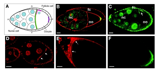
Localized distributions of grk, bcd, osk and
nos mRNAs. (A) Schematic showing grk (pink),
bcd (green) and osk (purple) mRNA localization in
mid-oogenesis (stage 9). nos mRNA is not yet localized at this stage.
The anteroposterior (AP) and dorsoventral (DV) axes are indicated. (B)
GFP-Stau (green), as proxy for osk mRNA, at the posterior pole of the
oocyte (oo) during mid-oogenesis. GFP-Stau is also detected in the nurse cell
(nc) cytoplasm. The actin cytoskeleton is highlighted in red with phalloidin.
fc, follicle cells. Orientation is the same as in A. (C-F)
Visualization of endogenous mRNAs using the MS2 system: (C) grk and
(D) bcd during mid-oogenesis; (E) bcd and (F) nos
in late oocytes. Owing to the promoter used, the MCP-GFP and MCP-RFP fusion
proteins are expressed in both the nurse cells and follicle cells, whereas the
MS2-tagged mRNAs are produced only in the nurse cells. MCP-GFP/RFP that is not
bound to mRNA enters both the nurse cell and follicle cell nuclei. Scale bars:
20 μm. Image in B was modified, with permission, from Huynh et al.
(Huynh et al., 2004); image in
C was modified, with permission, from Jaramillo et al.
(Jaramillo et al., 2008);
images in D and E are reproduced, with permission, from Weil et al.
(Weil et al., 2006). Image in
F is courtesy of K. Sinsimer (Princeton University, Princeton, NJ, USA).
bcd, bicoid; grk, gurken; GFP, green fluorescent protein;
MCP, MS2 coat protein; nos, nanos; osk, oskar; RFP, red
fluorescent protein.
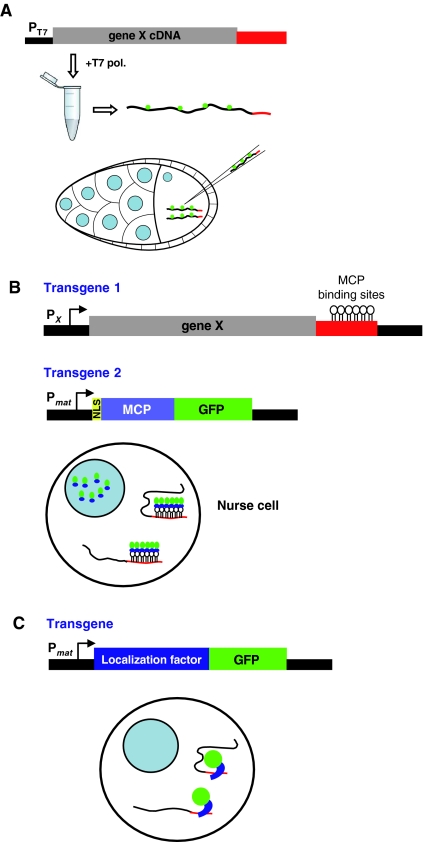
Fluorescent labeling methods. (A) A construct designed for in
vitro transcription of the gene of interest from a bacteriophage promoter (T7
in this example), with the coding region shown in gray and the 3′UTR
containing the RNA localization signal in red. Transcription of this construct
by T7 polymerase in the presence of a fluorophore-coupled nucleotide produces
fluorescently labeled RNA for injection into cultured egg chambers. RNA can be
injected directly into the oocyte as illustrated or into the nurse cells.
(B) In vivo labeling of endogenous mRNA by the MS2 system. This
strategy requires two components: a transgene (transgene 1) that encodes the
target RNA with an insertion of tandem copies of the stem-loop binding site
for the bacteriophage MS2 coat protein (MCP), shown here in the 3′ UTR,
usually under the control of its own promoter (Px); and a
transgene (transgene 2) that encodes a fluorescent protein fused to MCP (GFP
is shown here) under the control of a maternally active promoter
(Pmat). Transgenic fly lines for each component are
crossed together to generate females that express both the tagged RNA and the
MCP-GFP protein in their ovarian nurse cells. When the two transgenes are thus
coexpressed, the binding of MCP to its recognition motif labels the RNA with
GFP. The nuclear localization signal (NLS) in the MCP-GFP fusion protein
retains excess unbound protein in the nucleus, reducing cytoplasmic
background. Fluorescently labeled mRNA enters the oocyte from the nurse cells
(not shown). (C) Transgenic expression of GFP-tagged localization
factors. A transgene encoding a localization factor fused to a fluorescent
protein (e.g. GFP) under the control of its own or a maternally active
promoter. Expression of the transgene in the nurse cells of transgenic females
will result in production of the fusion protein in the nurse cells.
Colocalization of the fusion protein with the target RNA could occur in the
nurse cells or oocyte (not shown), through direct RNA-protein interaction (as
shown) or through their co-assembly into a larger RNP.
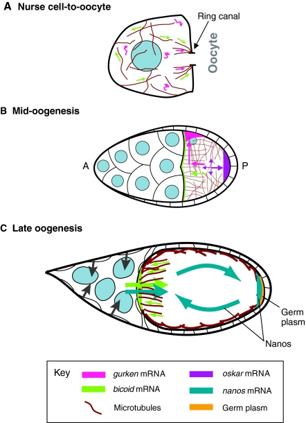
Models for mRNA localization. In all panels, microtubules are shown
in brown, nurse cell and follicle cell nuclei in blue. (A) Movements of
grk and bcd mRNAs within the nurse cells during
mid-oogenesis. Straight arrows indicate directed movement on microtubules,
squiggly arrows indicate movement of grk with cytoplasmic flows.
(B) Microtubule-dependent transport of grk, bcd and
osk mRNAs within the oocyte during mid-oogenesis. The oocyte nucleus
is shown in gray. Colored arrows show the directions of RNA movements.
(C) Localization of bcd and nos at late stages of
oogenesis. Contraction of the nurse cells for dumping is indicated by gray
arrows pointing inward; entry of bcd and nos into the oocyte
is indicated by large straight arrows. Small green arrows depict transport of
bcd on anterior microtubules, curved dark green arrows depict
diffusion of nos facilitated by ooplasmic streaming.
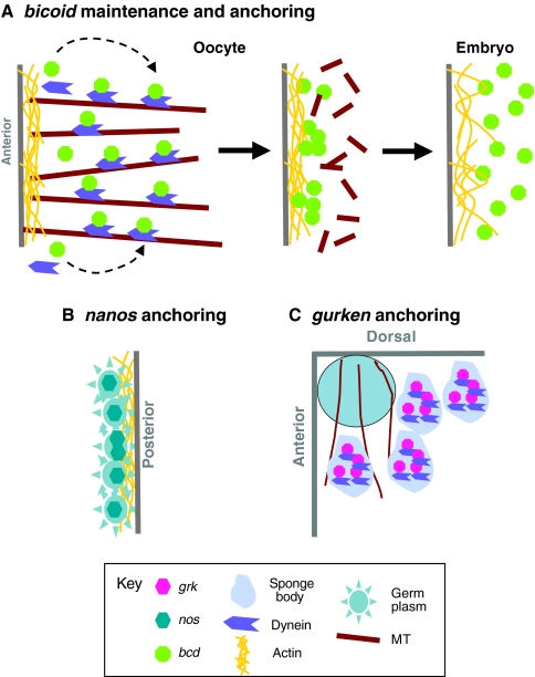
Maintenance of localized mRNAs. (A) Maintenance of
bcd mRNA at the anterior cortex of late oocytes by continual active
transport on microtubules (MT), transition to a static actin-dependent
anchoring mechanism at the end of oogenesis, and release from the tight
cortical anchor at fertilization. (B) Stable, actin-dependent anchoring
of nos mRNA and germ plasm (including osk) in late oocytes.
(C) Dynein-dependent anchoring of grk mRNA in sponge bodies
during mid-oogenesis. The oocyte nucleus is depicted as a blue circle.
References
-
- Ashraf, S. I., McLoon, A. L., Sclarsic, S. M. and Kunes, S. (2006). Synaptic protein synthesis associated with memory is regulated by the RISC pathway in Drosophila. Cell 124, 191-205. - PubMed
-
- Bertrand, E., Chartrand, P., Schaefer, M., Shenoy, S. M., Singer, R. H. and Long, R. M. (1998). Localization of ASH1 mRNA particles in living yeast. Mol. Cell 2, 437-445. - PubMed
-
- Breitwieser, W., Markussen, F. H., Horstmann, H. and Ephrussi, A. (1996). Oskar protein interaction with Vasa represents an essential step in polar granule assembly. Genes Dev. 10, 2179-2188. - PubMed
Publication types
MeSH terms
Substances
Grants and funding
LinkOut - more resources
Full Text Sources
Other Literature Sources
Molecular Biology Databases
