. 2009 Sep;297(3):H1032-9.
doi: 10.1152/ajpheart.00374.2009. Epub 2009 Jul 10.

A(1) adenosine receptor-mediated PKC and p42/p44 MAPK signaling in mouse coronary artery smooth muscle cells

Affiliations

A(1) adenosine receptor-mediated PKC and p42/p44 MAPK signaling in mouse coronary artery smooth muscle cells

Habib R Ansari et al. Am J Physiol Heart Circ Physiol. 2009 Sep.

Abstract

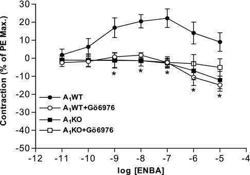
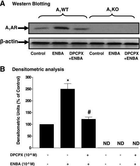
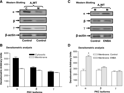
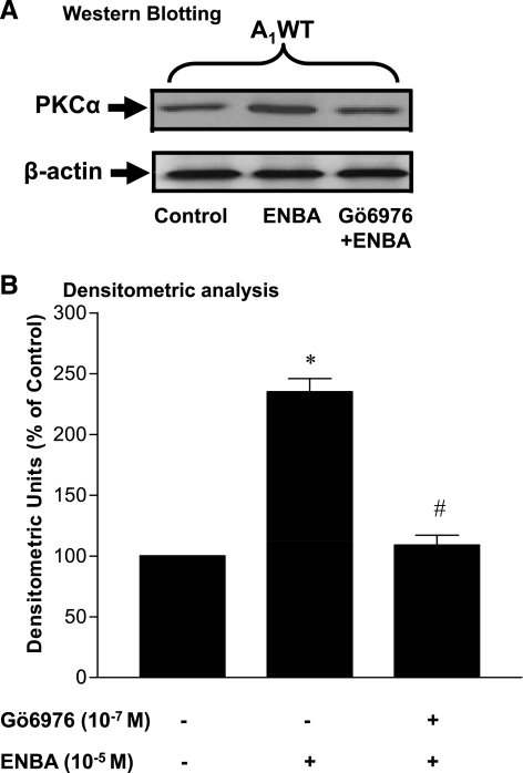
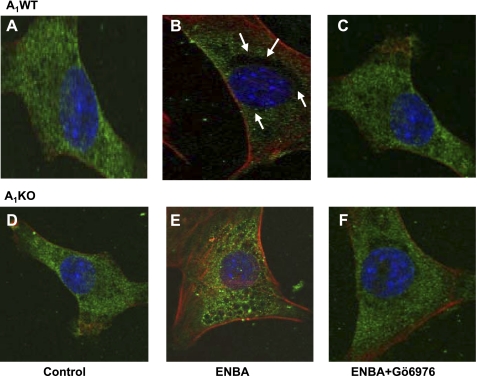
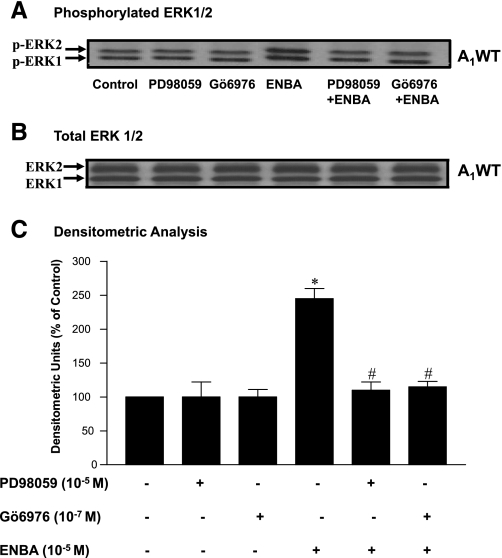
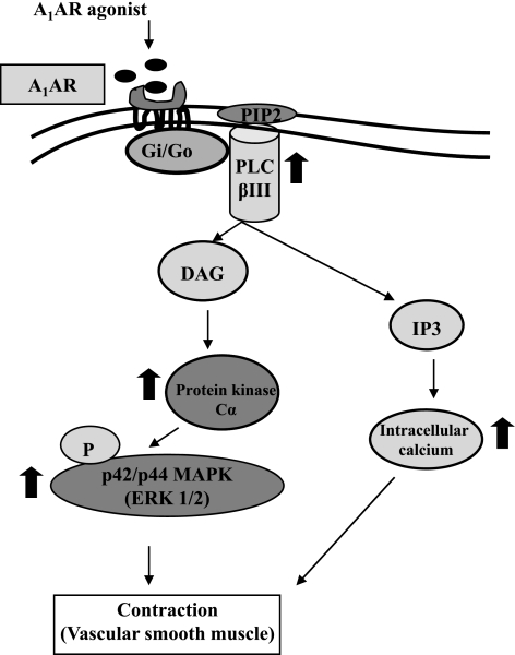
The A(1) adenosine receptor (A(1)AR) is coupled to G(i)/G(o) proteins, but the downstream signaling pathways in smooth muscle cells are unclear. This study was performed in coronary artery smooth muscle cells (CASMCs) isolated from the mouse heart [A(1)AR wild type (A(1)WT) and A(1)AR knockout (A(1)KO)] to delineate A(1)AR signaling through the PKC pathway. In A(1)WT cells, treatment with (2S)-N(6)-(2-endo-norbornyl)adenosine (ENBA; 10(-5)M) increased A(1)AR expression by 150%, which was inhibited significantly by the A(1)AR antagonist 1,3-dipropyl-8-cyclopentylxanthine (10(-6)M), but not in A(1)KO CASMCs. PKC isoforms were identified by Western blot analysis in the cytosolic and membrane fractions of cell homogenates of CASMCs. In A(1)WT and A(1)KO cells, significant levels of basal PKC-alpha were detected in the cytosolic fraction. Treatment with the A(1)AR agonist ENBA (10(-5)M) translocated PKC-alpha from the cytosolic to membrane fraction significantly in A(1)WT but not A(1)KO cells. Phospholipase C isoforms (betaI, betaIII, and gamma(1)) were analyzed using specific antibodies where ENBA treatment led to the increased expression of PLC-betaIII in A(1)WT CASMCs while having no effect in A(1)KO CASMCs. In A(1)WT cells, ENBA increased PKC-alpha expression and p42/p44 MAPK (ERK1/2) phospohorylation by 135% and 145%, respectively. These effects of ENBA were blocked by Gö-6976 (PKC-alpha inhibitor) and PD-98059 (p42/p44 MAPK inhibitor). We conclude that A(1)AR stimulation by ENBA activates the PKC-alpha signaling pathway, leading to p42/p44 MAPK phosphorylation in CASMCs.


Figures

Fig. 1.
Fig. 1.
Effect of the PKC-α inhibitor Gö-6976 on (2S)-N6-(2-endo-norbornyl) adenosine (ENBA)-induced contractile responses in A1 adenosine receptor (A1AR) wild-type (A1WT) and A1AR knockout (A1KO) mouse mesenteric arteries. PE, phenylephine. Results are expressed as means ± SE; n = 4. *P < 0.05 compared with A1WT (untreated) controls.
Fig. 2.
Fig. 2.
A1AR protein expression in A1WT and A1KO coronary artery smooth muscle cells (CASMCs). A: Western blot analysis. DPCPX, 1,3-dipropyl-8-cyclopentylxanthine. B: densitometric analysis. ND, not detected. Data are presented as means ± SE; n = 5–7. *P < 0.05 compared with A1WT (untreated) controls; #P < 0.05 compared with ENBA alone.
Fig. 3.
Fig. 3.
Phospholipase C (PLC) isoform (βI, βIII, and γ1) expression in A1WT CASMCs. A: Western blot analysis for PLC isoforms. B: densitometric analysis. data are presented as means ± SE; n = 3–4. *P < 0.05 compared with the respective controls.
Fig. 4.
Fig. 4.
Distribution of PKC isoforms (α, β, and γ) in the cytosolic fraction (C) and membrane fraction (M) with and without ENBA treatment of A1WT CASMCs. A and C: Western blots analysis. B and D: densitometric analysis. Data are presented as means ± SE; n = 4–5. *P < 0.05 compared with the respective membrane fractions.
Fig. 5.
Fig. 5.
Effect of Gö-6976 (PKC-α inhibitor) on ENBA-induced PKC-α expression by Western blot analysis in A1WT CASMCs. A: Western blot analysis. B: densitometric analysis. Data are presented as means ± SE; n = 6. *P < 0.05 compared with the untreated control in A1WT CASMCs; #P < 0.05 compared with ENBA alone in A1WT CASMCs.
Fig. 6.
Fig. 6.
PKC-α immunoreactivity in A1WT and A1KO CASMCs. A and D: representative immunofluorescence photomicrographs of control cells showing PKC-α with green staining in A1WT (A) and A1KO (D) CASMCs. Nuclei were stained with 4′,6-diamidino-2-phenylindole (blue). The green color around the nucleus shows PKC-α immunoreactivity. Arrows show the translocation of PKC-α in A1WT CASMCs toward the membrane after ENBA treatment (black empty area around the nucleus; B). ENBA-induced translocation toward the membrane was blocked by the PKC-α inhibitor Gö-6976 (C). Membrane translocation was not seen after ENBA treatment in A1KO CASMCs (E and F).
Fig. 7.
Fig. 7.
Effect of PD-98059 (MAPK inhibitor) and Gö-6976 (PKC-α inhibitor) on ENBA-induced p42/p44 MAPK (ERK1/2) phosphorylation in A1WT CASMCs. A: phosphorylated p42/p44 MAPK (p-ERK1/2). B: total ERK. C: densitometric analysis of p42/p44 MAPK (ERK1/2) phosphorylation. Data are presented as means ± SE; n = 6. *P < 0.05 compared with controls (untreated); #P < 0.05 compared with ENBA alone.
Fig. 8.
Fig. 8.
Putative signaling mechanism for A1AR-mediated contraction in vascular smooth muscle. The activation of the A1AR originates from the binding of an extracellular ligand (A1AR agonist) and is followed by the activation of a G protein on the cytosolic side of the plasma membrane. The G protein, using GTP as an energy source, then activates PLC-βIII, leading to the activation of PKC-α via diacylglycerol (DAG). PKC-α then phosphorylates p42/p44 MAPK, leading to the contraction of smooth muscle. On the other hand, inositol (1,4,5)-trisphosphate (IP3) also causes the contraction of smooth muscle through an increase in intracellular calcium. PIP2: phosphatidylinositol bisphosphate.

References

    1. Ansari HR, Husain S, Abdel-Latif AA. Activation of p42/p44 mitogen-activated protein kinase and contraction by prostaglandin F2alpha, ionomycin, and thapsigargin in cat iris sphincter smooth muscle: inhibition by PD98059, KN-93, and isoproterenol. J Pharmacol Exp Ther 299: 178–186, 2001. - PubMed
    1. Ansari HR, Nadeem A, Tilley SL, Mustafa SJ. Involvement of COX-1 in A3 adenosine receptor-mediated contraction through endothelium in mice aorta. Am J Physiol Heart Circ Physiol 293: H3448–H3455, 2007. - PubMed
    1. Bradford M. A rapid and sensitive method for the quantitation of microgram quantities of protein utilizing the principle of protein-dye binding. Anal Biochem 72: 248–254, 1976. - PubMed
    1. Ding Y, Schwartz D, Posner P, Zhong J. Hypotonic swelling stimulates L-type Ca2+ channel activity in vascular smooth muscle cells through PKC. Am J Physiol Cell Physiol 287: C413–C421, 2004. - PubMed
    1. Fredholm BB, Ijzerman AP, Jacobson KA, Klotz KN, Linden J. International Union of Pharmacology: XXV. Nomenclature and classification of adenosine receptors. Pharmacol Rev 53: 527–552, 2001. - PMC - PubMed

Publication types

MeSH terms

LinkOut - more resources