. 2009 Aug 1;183(3):1732-8.
doi: 10.4049/jimmunol.0802923. Epub 2009 Jul 10.

IL-6 is required for airway mucus production induced by inhaled fungal allergens

Affiliations

IL-6 is required for airway mucus production induced by inhaled fungal allergens

Wendy A Neveu et al. J Immunol. .

Abstract

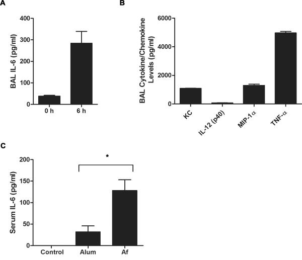
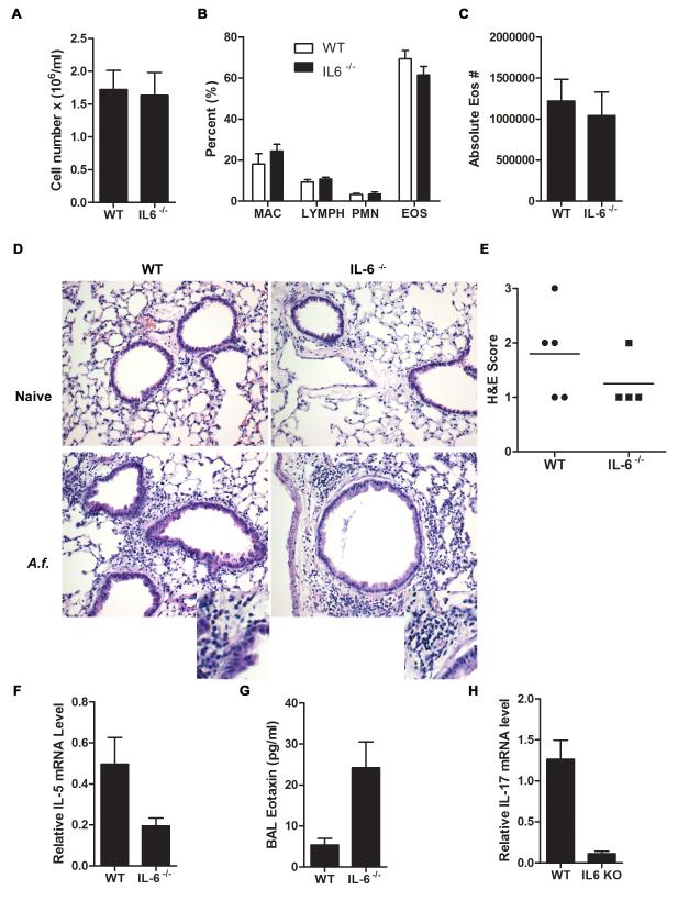
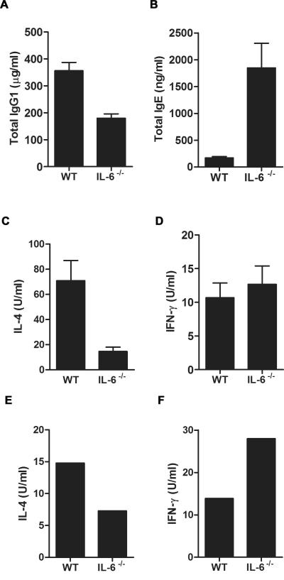
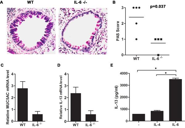
Allergic asthma is caused by inhaled allergens and is characterized by airway eosinophilia, as well as mucus hypersecretion, which can lead to airflow obstruction. Despite the association of increased IL-6 levels with human atopic asthma, the contribution of IL-6 to the development of allergic airway inflammation triggered by inhaled allergens remains unclear. In this study, we examined the role of IL-6 in a mouse model of allergic airway inflammation induced by direct airway exposure to extracts of Aspergillus fumigatus, a common allergen in humans. We show that inhaled A. fumigatus extracts rapidly trigger the production of IL-6 in the airways. IL-6 appears to be dispensable for the recruitment of eosinophils to the lung during the development of allergic airway inflammation. However, IL-6 is essential for mucus hypersecretion by airway epithelial cells triggered in response to inhaled A. fumigatus Ags. Impaired mucus production caused by IL-6 deficiency correlates with a severe reduction in the levels of IL-13, a major inducer of mucin glycoproteins. Thus, IL-6 is a key regulator of specific hallmark features of allergic airway inflammation and it could be a potential target for pulmonary diseases that are associated with goblet cell metaplasia and mucus hypersecretion.


Figures

Figure 1
Figure 1
A.fumigatus induces rapid IL-6 production in the airway and peripheral blood. (A) Wild type mice (n=4) were o.p. administered A.f. extracts. IL-6 levels in the BALF were assessed at 0 and 6 h post exposure by ELISA. The results indicate the mean ± SEM (p<0.05). (B) Wild type mice were o.p. administered A.f. extracts and 6 h later cytokine/chemokine levels in BALF were determined by Bio-Plex. (C) Wild type mice were i.p. administered OVA with alum (n=5) or A.f. (n=4) and 6 h later serum IL-6 levels were measured by ELISA (*, p<0.05). Data are representative of two or three independent experiments.
Figure 2
Figure 2
IL-6 does not contribute to eosinophilic infiltration in the lung. (A-C) Wild type and IL-6−/− mice (n=4) were administered A.f. extracts o.p. on day 0, 7, and 14. BALF was collected 48 h after last A.f. dose. (A) Total cell count in BALF was determined (p=0.89). (B) Relative percentage of macrophages (MAC), lymphocytes (LYMPH), neutrophils (PMN), and eosinophils (EOS) in BALF (p>0.05). (C) Absolute number of eosinophils in BALF (p=0.89). (D) H&E staining of paraffin embedded lungs sections from unexposed (naïve) and A.f.-exposed wild type and IL-6−/− mice (×200) as described in (A). Insets show expanded image. (E) Quantification of lung inflammation in A.f.-exposed wild type (n=5) and IL-6−/− (n=4) mice as indicated by H&E staining (p=0.34). (F) IL-5 expression in total lung tissue from A.f.-exposed wild type (n=5) and IL-6−/− (n=4) mice was determined by real time RT-PCR and normalized to 18S RNA (p<0.05). (G) BALF eotaxin levels from A.f.-exposed wild type (n=9) and IL-6−/− (n=8) mice were quantified by ELISA (p<0.05). (H) Wild type (n=5) and IL-6−/− (n=4) mice were exposed to A.f. extracts as described in (A) and IL-17 expression in CD4+ T cells purified from wild type and IL-6−/− mice was determined by real time RT-PCR and normalized to β2m RNA (p<0.05). Data are representative of two or three independent experiments.
Figure 3
Figure 3
IL-6 is required for IgG1, but negatively regulates IgE production in A.f.-induced allergic airway inflammation. (A) and (B) Wild type and IL-6−/− mice (n=4) were exposed to A.f. extracts as described in Fig. 2. (A) Serum IgG1 (p<0.05) and (B) IgE levels (p<0.05) were determined by ELISA. (C) and (D) Splenic CD4+ T cells from A.f.-exposed wild type (n=7) and IL-6−/− (n=4) mice were restimulated ex vivo for 24 h with anti-CD3 (5 μg/ml) and anti-CD28 (1 μg/ml) mAbs. Cell supernatant was analyzed for (C) IL-4 (p<0.05) and (D) IFNγ (p=0.51) by ELISA. (E) and (F) FACS-sorted lung CD4+ T cells pooled from three wild type mice and three IL-6−/− mice exposed to A.f. extracts were restimulated ex vivo with A.f. (5 μg/ml) for 72h. Cell supernatant was analyzed for (E) IL-4 and (F) IFNγ by ELISA. Data are representative of two or three independent experiments.
Figure 4
Figure 4
IL-6 is necessary for IL-21 production by CD4+ T cells during allergic airway inflammation. (A) IL-21 expression levels in CD4+ T cells from A.f.-exposed wild type (n=5) and IL-6−/− (n=4) mice were examined by real time RT-PCR (p<0.05). (B) Serum IgE levels were analyzed in non-exposed wild type (n=5), IL-21−/− (n=7), and IL-6−/− (n=5) mice (*, p<0.05). Data are representative of two or three independent experiments.
Figure 5
Figure 5
IL-6 mediates mucus production by lung epithelium through the induction of IL-13 in CD4+ T cells in allergic airway inflammation. (A-D) Wild type (n=5) and IL-6−/− (n=4) mice were exposed to A.f. extracts as described in Fig. 2. (A) Mucus production in the lung was visualized by PAS staining of paraffin embedded lung sections. Photomicrographs from A.f.-challenged wild type (left) and IL-6−/− mice (right) are original magnification, ×200. (B) Histological score indicating intensity of PAS staining for each group (p<0.05). (C) Muc5AC (p<0.05) and (D) IL-13 (p<0.05) expression in the lung were determined by real time RT-PCR and normalized to 18S RNA. (E) Naïve CD4+ T cells from wild type mice were activated in vitro with anti-CD3 and anti-CD28 mAbs in the presence or absence of IL-6 (100 ng/ml) or IL-4 (1×10³ U/ml). IL-13 was measured in the culture supernatant after 48 h by ELISA in triplicate (*, p<0.05 CD4+ T cells treated with IL-6 compared with cells activated in the presence of medium alone or IL-4).

References

    1. Kishimoto T. Interleukin-6: from basic science to medicine--40 years in immunology. Annu Rev Immunol. 2005;23:1–21. - PubMed
    1. Diehl S, Anguita J, Hoffmeyer A, Zapton T, Ihle JN, Fikrig E, Rincon M. Inhibition of Th1 differentiation by IL-6 is mediated by SOCS1. Immunity. 2000;13:804–815. - PubMed
    1. Diehl S, Chow CW, Weiss L, Palmetshofer A, Twardzik T, Rounds L, Serfling E, Davis RJ, Anguita J, Rincon M. Induction of NFATc2 expression by interleukin 6 promotes T helper type 2 differentiation. J. Exp. Med. 2002;196:39–49. - PMC - PubMed
    1. Yang Y, Ochando J, Yopp A, Bromberg JS, Ding Y. IL-6 plays a unique role in initiating c-Maf expression during early stage of CD4 T cell activation. J Immunol. 2005;174:2720–2729. - PubMed
    1. Veldhoen M, Hocking RJ, Atkins CJ, Locksley RM, Stockinger B. TGFbeta in the context of an inflammatory cytokine milieu supports de novo differentiation of IL-17-producing T cells. Immunity. 2006;24:179–189. - PubMed

Publication types