. 2009 Jul;5(7):e1000559.
doi: 10.1371/journal.pgen.1000559. Epub 2009 Jul 10.

Specific loss of histone H3 lysine 9 trimethylation and HP1gamma/cohesin binding at D4Z4 repeats is associated with facioscapulohumeral dystrophy (FSHD)

Affiliations

Specific loss of histone H3 lysine 9 trimethylation and HP1gamma/cohesin binding at D4Z4 repeats is associated with facioscapulohumeral dystrophy (FSHD)

Weihua Zeng et al. PLoS Genet. 2009 Jul.

Abstract

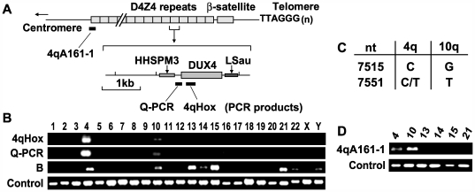
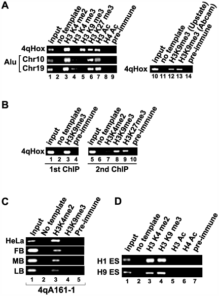
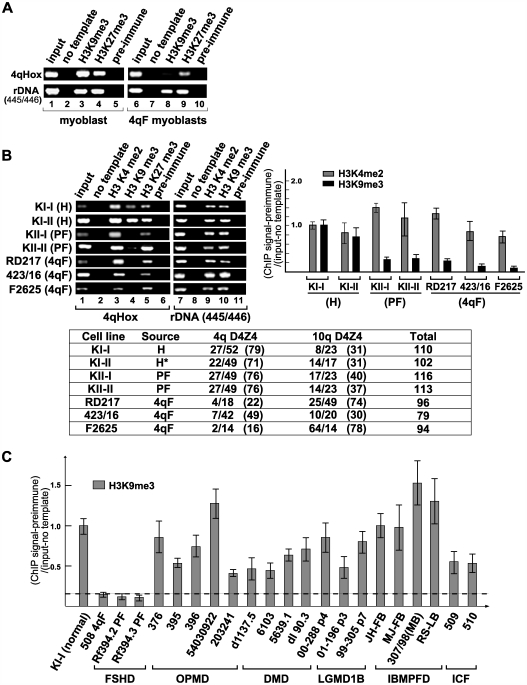
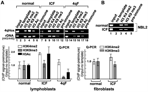
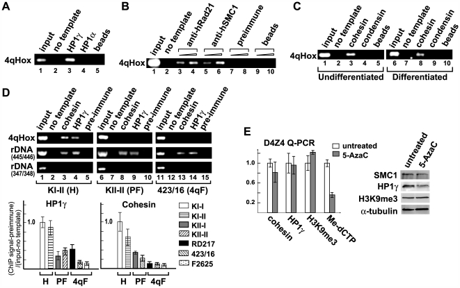
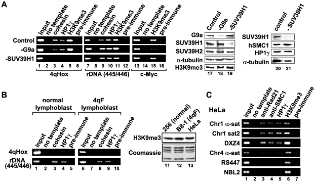
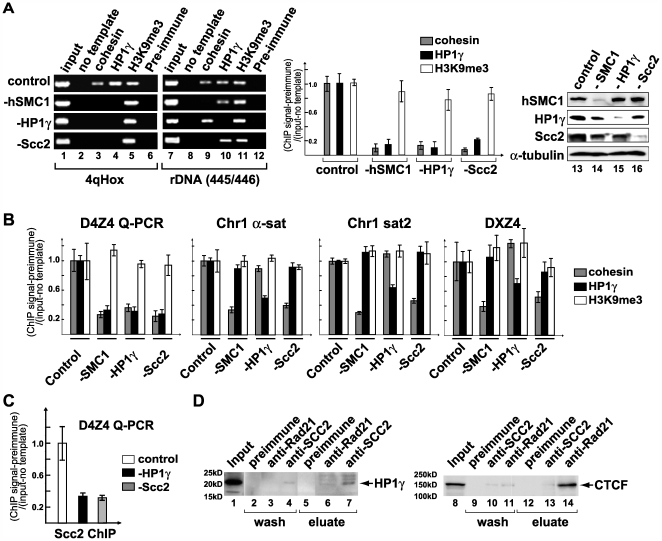
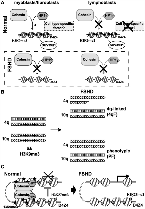
Facioscapulohumeral dystrophy (FSHD) is an autosomal dominant muscular dystrophy in which no mutation of pathogenic gene(s) has been identified. Instead, the disease is, in most cases, genetically linked to a contraction in the number of 3.3 kb D4Z4 repeats on chromosome 4q. How contraction of the 4qter D4Z4 repeats causes muscular dystrophy is not understood. In addition, a smaller group of FSHD cases are not associated with D4Z4 repeat contraction (termed "phenotypic" FSHD), and their etiology remains undefined. We carried out chromatin immunoprecipitation analysis using D4Z4-specific PCR primers to examine the D4Z4 chromatin structure in normal and patient cells as well as in small interfering RNA (siRNA)-treated cells. We found that SUV39H1-mediated H3K9 trimethylation at D4Z4 seen in normal cells is lost in FSHD. Furthermore, the loss of this histone modification occurs not only at the contracted 4q D4Z4 allele, but also at the genetically intact D4Z4 alleles on both chromosomes 4q and 10q, providing the first evidence that the genetic change (contraction) of one 4qD4Z4 allele spreads its effect to other genomic regions. Importantly, this epigenetic change was also observed in the phenotypic FSHD cases with no D4Z4 contraction, but not in other types of muscular dystrophies tested. We found that HP1gamma and cohesin are co-recruited to D4Z4 in an H3K9me3-dependent and cell type-specific manner, which is disrupted in FSHD. The results indicate that cohesin plays an active role in HP1 recruitment and is involved in cell type-specific D4Z4 chromatin regulation. Taken together, we identified the loss of both histone H3K9 trimethylation and HP1gamma/cohesin binding at D4Z4 to be a faithful marker for the FSHD phenotype. Based on these results, we propose a new model in which the epigenetic change initiated at 4q D4Z4 spreads its effect to other genomic regions, which compromises muscle-specific gene regulation leading to FSHD pathogenesis.


Conflict of interest statement

The authors have declared that no competing interests exist.

Figures

Figure 1
Figure 1. Specific PCR amplification of D4Z4 repeat sequences.
(A) A schematic diagram of the 4qter D4Z4 repeat region and a single D4Z4 repeat. PCR products for Q-PCR and 4qHox primer pairs are indicated by black bars. The DUX4 ORF and a GC-rich sequence homologous to the low-copy repeat HHSPM3 are shown. (B) PCR analysis of a DNA mapping panel consisting of genomic DNA isolated from mouse and hamster somatic cell hybrids containing individual human chromosomes using the 4qHox and Q–PCR primer pairs. The “B” PCR primer pair also binds to a region within D4Z4. However, it amplified not only chromosomes 4 and 10, but also several other chromosomes presumably due to crossreactivity to other D4Z4-like repeat sequences, and therefore, was not used for the experiments. For control PCR reactions, primers corresponding to the mouse β-globin locus were used for the chromosome 1, 16, 17, 20, and 21 hybrids, while primers for hamster rDNA regions were utilized for the other hybrids. PCR analysis of additional mouse somatic cell hybrids for human chromosomes 4, 10, 13, 14, 15, and 21 also yielded similar results (data not shown). (C) Sequence polymorphisms between 4q and 10q D4Z4 ,. The nucleotide positions (nt) of the sequence polymorphisms are based on AF117653 in the GenBank/EMBL Nucleotide Sequence Database. (D) PCR analysis using the 4qA161-1 primer pair against genomic DNA from mouse somatic cell hybrids containing human chromosomes 4, 10, 13, 14, 15, and 21.
Figure 2
Figure 2. D4Z4 chromatin contains both euchromatic and heterochromatic histone modifications.
(A) Antibodies specific for H3K4me2, H3K4me3, H3K9me3, H3K27me3, H3Ac, and acetylated H4 (H4Ac), as well as control preimmune IgG, were used for ChIP in HeLa cells. The ChIP DNA was amplified using 4qHox primers and primers specific for regions on chromosomes 10 and 19 containing short Alu repeat sequences. The presence of H3K9me3 was confirmed by two different antibodies (lanes 10–14) . (B) Double-ChIP analysis of D4Z4 histone modifications. H3K9me3 ChIP (1st ChIP) was eluted and followed by the second (2nd) ChIP reactions using antibodies specific for H3K4me2, H3K9me3, H3K27me3, or preimmune IgG. The ChIP DNA was amplified using 4qHox primers. (C) The proximal region of the D4Z4 cluster is euchromatic. ChIP analysis of the first proximal D4Z4 repeat using the 4qA161-1 primer pair (See Figure 1D for sequence amplification specificity) was performed in HeLa, normal human fibroblasts (FB), myoblasts (MB), and lymphoblasts (LB). (D) Histone ChIP in human ES cells. ChIP DNA derived from the ES cell lines H1 and H9 was amplified by 4qHox primers. Antibodies used for ChIP are indicated at the top.
Figure 3
Figure 3. Histone H3 lysine 9 trimethylation is specifically lost at D4Z4 in both 4q-linked and phenotypic FSHD.
(A) ChIP analysis of H3K9me3 and H3K27me3 at D4Z4 in normal and FSHD (4qF) myoblasts. The rDNA region (445/446) serves as a positive control. (B) H3K9me3 is specifically lost in FSHD fibroblasts. Endpoint PCR analysis with 4qHox primers by agarose gel electrophoresis and quantitation of real-time PCR with Q-PCR primers are shown. The rDNA region, which was positive for HP1 and cohesin binding, was used for comparison (445/446) (See Figure 5). PCR signals were normalized with preimmune, input, and no template PCR signals. Primary cells derived from healthy (normal) (H), phenotypic (PF), and 4q-linked (4qF) individuals were analyzed as indicated. D4Z4 repeat numbers for 4q and 10q alleles as well as the total D4Z4 repeat numbers are shown in the table. The asterisk indicates a clinically unaffected individual with DNA hypomethylation at D4Z4, whose two offspring developed phenotypic FSHD (KII-I and KII-II). (C) H3K9me3 ChIP analysis of different muscular dystrophy patient cells. The graph contains one 4qF (508) and two PF (Rf394.2 and Rf394.3) patient fibroblast samples, five OPMD patient fibroblast samples carrying alanine repeat insertions in the PABPN1 gene (376, 395, 396, 54030922, and 203241), four DMD patient fibroblast samples with mutations in the dystrophin gene (d1137.5, 6103, 5639.1, and dl90.3), three LGMD patient fibroblast samples with heterozygous mutations in the LMN gene (00–288, 01–196, 99–305) ,, two ICF patient fibroblast samples , and four IBMPFD patient samples (two fibroblast and two lymphoblast) with mutations in the VCP gene (JH-FIB, MJ-FIB, 307/98, and RS-LCL) . The KI-I (normal) fibroblast sample serves as a control. H3K9me3 was also retained in two additional control fibroblast samples (302, 557/96) (data not shown).
Figure 4
Figure 4. H3K9me3 at D4Z4 is maintained in ICF patient cells.
(A) ChIP analysis by endpoint PCR using 4qHox primers was performed using normal, ICF, and 4q-linked FSHD (4qF) lymphoblasts with antibodies specific for H3K4me2, H3K9me3, and H3Ac, and preimmune IgG as indicated at the top. The rDNA region (445/446) serves as a positive control. ChIP analysis by real-time PCR using Q-PCR primers for H3K4me2, H3K9me3, and H3Ac is also shown. Similar results were obtained with ICF fibroblasts (Q-PCR results are shown at the bottom right). (B) H3K9me3 is intact at the NBL2 repeat region in ICF cells. Similar ChIP analysis was performed using PCR primers specific for the NBL2 repeat sequence.
Figure 5
Figure 5. The binding of HP1γ and cohesin to D4Z4 is lost in FSHD patients.
(A) HP1γ, but not HP1α, binds to D4Z4 in HeLa cells. 4qHox endpoint PCR of ChIP DNA using antibodies against HP1γ and HP1α is shown. Immunoprecipitation with protein A beads alone serves as a negative control. (B) Comparison of ChIP analyses using antibodies specific for two different subunits of cohesin (hSMC1 and hRad21). Preimmune IgG and protein A beads alone were used as negative controls. Two different amounts of ChIP DNA were used for endpoint PCR with 4qHox primers as indicated. The remainder of the cohesin ChIP experiments were carried out using anti-hRad21 antibody. (C) Cohesin binds to the 4qHox region in undifferentiated and differentiated primary human myoblasts. Cohesin ChIP was compared to that of condensin, another major SMC-containing complex, and protein A beads control. (D) ChIP PCR analyses using 4qHox primers of HP1γ and cohesin binding in H, PF and 4qF fibroblasts as in Figure 3B. Representative samples of the 4qHox PCR products on an agarose gel are shown. PCR primers corresponding to the rDNA locus serve as positive (445/446) and negative (347/348) controls for HP1 and cohesin binding. Real-time PCR analysis using Q-PCR primers of HP1γ and cohesin ChIP is shown underneath. A similar loss of HP1γ and cohesin was also observed in 4qF myoblasts (data not shown). (E) The effect of DNA hypomethylation on cohesin and HP1γ binding and H3K9me3. HeLa cells were treated with 5-AzaC and ChIP-PCR assays were performed using antibodies specific for Rad21 (“cohesin”), HP1γ and H3K9me3 and Q–PCR primers specific for D4Z4. Hypomethylation of DNA was confirmed by MeCIP using antibody specific for 5-methylcytidine. The ChIP and MeCIP signal intensity was normalized by genomic DNA input control and pre-immune control. No significant decrease of cohesin and HP1γ binding and H3K9me3 was observed. Western analysis of cohesin, HP1γ and H3K9me3 levels in untreated and 5-AzaC-treated cells is also shown.
Figure 6
Figure 6. SUV39H1 HMTase is solely responsible for H3K9me3 at D4Z4, which is necessary, but not sufficient, for the recruitment of HP1γ and cohesin.
(A) SUV39H1 is responsible for H3K9me3 and HP1γ/cohesin association at D4Z4. HeLa cells were treated with siRNA specific for SUV39H1, G9a, or control siRNA, and ChIP analysis using 4qHox primers was performed for the presence of cohesin, HP1γ and H3K9me3 (lanes 1–16). Preimmune IgG serves as a negative control. The rDNA (445/446) and c-Myc regions were used for comparison. Western-blot analysis of G9a and SUV39H1 siRNA depletion is also shown (lanes 17–21). Depleted proteins are indicated at the top and proteins detected by western blot analysis are indicated on the left. α-tubulin serves as a loading control. (B) HP1γ and cohesin binding to D4Z4 is cell type-specific. ChIP analysis of D4Z4 and rDNA regions was performed using normal and 4qF lymphoblasts (lanes 1–10). Western blot analysis comparing the level of H3K9me3 between HeLa and lymphoblasts (256 (normal) and B8-1 (4qF)) is also shown (lanes 11–13). Coomassie staining of core histones is included as a loading control. (C) Not all H3K9me3-positive repeats are bound by HP1γ and cohesin. Six different repeat sequences (as in Figure S1) were tested for cohesin and HP1γ binding in HeLa cells. While H3K9me3 was detected at all six repeat sequences tested, cohesin and HP1γ binding was found at only three repeats (α-sat and sat2 on chromosome 1 and DXZ4).
Figure 7
Figure 7. D4Z4-specific co-recruitment of HP1γ, cohesin, and cohesin loading factor Scc2.
(A) Binding of HP1γ and cohesin to D4Z4 is interdependent. ChIP analysis of HeLa cells after individual depletion of the cohesin subunit hSMC1, HP1γ, or the cohesin loading factor Scc2 by siRNA as indicated (lanes 1–12). Cohesin and HP1γ binding was compared between D4Z4 and rDNA (445/446). Real-time PCR analysis using Q–PCR primers is shown underneath. Western blot analysis of hSMC1, HP1γ and Scc2 depletion is also shown (lanes 13–16). (B) HP1γ and cohesin binding do not affect each other at other repeat sequences. Realtime PCR analysis of Rad21 (“cohesin”), HP1γ and H3K9me3 ChIP DNA from HeLa cells treated with control, SMC1, HP1γ, or Scc2 siRNA as indicated using Q-PCR primers specific for D4Z4, α-sat and sat2 repeat sequences on chromosome 1, and DXZ4 (as in Figure S2). (C) Scc2 binding to D4Z4 is compromised by HP1γ depletion. Realtime PCR analysis of Scc2 ChIP DNA from HeLa cells treated with control, HP1γ, or Scc2 siRNA as indicated using Q-PCR primers specific for D4Z4. (D) Coimmunoprecipitation (co-IP)–western blot analysis of cohesin and Scc2 interaction with HP1γ. HeLa nuclear extracts were used for co-IP using antibody specific for Scc2 or cohesin (Rad21) as previously described ,. After low-salt washes, precipitated materials were eluted with 1.0 M KCl (“wash”) and further eluted with 2.0 M guanidine-HCl (“eluate”). Eluted proteins were analyzed by SDSPAGE and western blotting using antibody specific for HP1γ. For comparison, a similar co-IP analysis was performed and probed with antibody specific for CTCF.
Figure 8
Figure 8. Schematic models of chromatin changes and the possible consequences in FSHD.
(A) Schematic summary of the cell type-specific chromatin assembly at D4Z4 and its loss in FSHD. HP1γ and cohesin are co-recruited to D4Z4 that harbors SUV39H1-dependent H3K9me3 in certain cell types, including myoblasts and fibroblasts. In lymphoblasts, however, despite the presence of H3K9me3, HP1γ and cohesin fail to associate with D4Z4 raising the possibility that HP1γ and cohesin are involved in cell type-specific chromatin organization and that a putative cell type-specific factor(s) (or modification(s)) required for their recruitment may not be present in lymphoblasts. Thus, while loss of H3K9me3 at D4Z4 in FSHD has no consequence at D4Z4 in lymphoblasts, it leads to abolishment of HP1γ/cohesin binding in myoblasts, resulting in a detrimental effect on chromatin organization leading to muscular dystrophy. (B) Coordinated loss of H3K9me3 on 4q and 10q D4Z4 in 4qF and PF. H3K9me3 (shown by black triangles) clustered in the subdomains of D4Z4 repeat regions (distribution hypothetical) in normal cells is lost in both types of FSHD. (C) A possible model for the spreading of the epigenetic change at D4Z4 to other genomic regions in FSHD. HP1γ and cohesin may contribute to the physical interactions of the heterochromatic D4Z4 region with other genomic regions leading to the spreading of the silencing effect to putative target genes in normal cells. In FSHD, the loss of H3K9me3 (but not H3K27me3), HP1γ, and cohesin from D4Z4 results in loss of chromatin interaction and derepression of these genes leading to muscular dystrophy.

References

    1. Padberg GW. Facioscapulohumeral disease. [MD Thesis] Leiden, The Netherlands: Leiden University; 1982.
    1. van der Maarel SM, Frants RR. The D4Z4 repeat-mediated pathogenesis of facioscapulohumeral muscular dystrophy. Am J Hum Genet. 2005;76:375–386. - PMC - PubMed
    1. Gabellini D, Green MR, Tupler R. Inappropriate gene activation in FSHD: a repressor complex binds a chromosomal repeat deleted in dystrophic muscle. Cell. 2002;110:339–348. - PubMed
    1. Gabellini D, D'Antona G, Moggio M, Prelle A, Zecca C, et al. Facioscapulohumeral muscular dystrophy in mice overexpressing FRG1. Nature. 2006;439:973–977. - PubMed
    1. Winokur ST, Chen YW, Masny PS, Martin JH, Ehmsen JT, et al. Expression profiling of FSHD muscle supports a defect in specific stages of myogenic differentiation. Hum Mol Genet. 2003;12:2895–2907. - PubMed

Publication types

MeSH terms