. 2010 May;121(1):65-78.
doi: 10.1007/s10549-009-0457-3. Epub 2009 Jul 11.

Antimitotic chemotherapeutics promote adhesive responses in detached and circulating tumor cells

Affiliations

Antimitotic chemotherapeutics promote adhesive responses in detached and circulating tumor cells

Eric M Balzer et al. Breast Cancer Res Treat. 2010 May.

Abstract

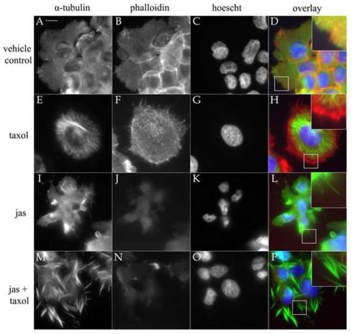
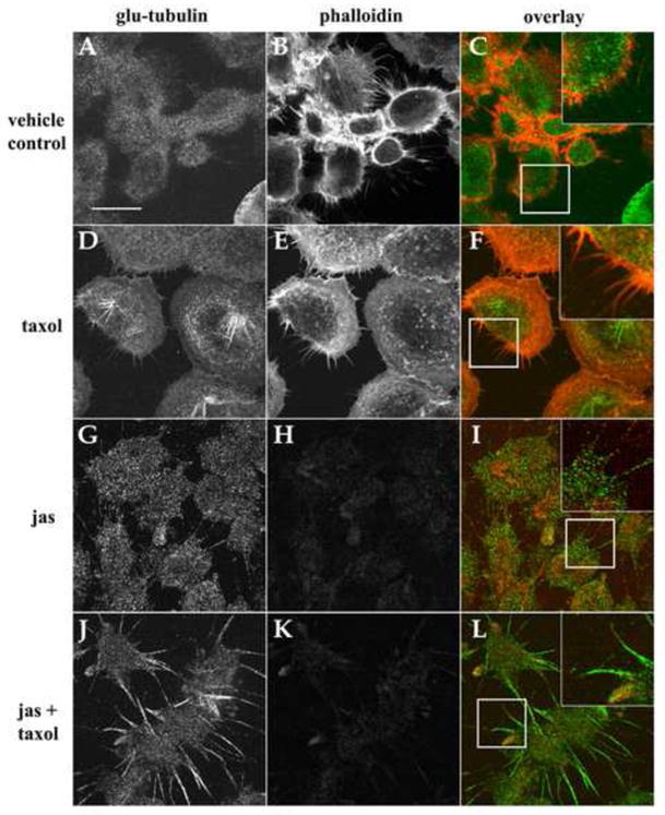
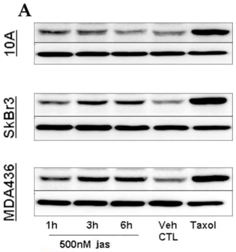
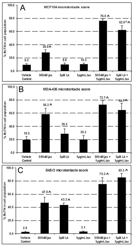
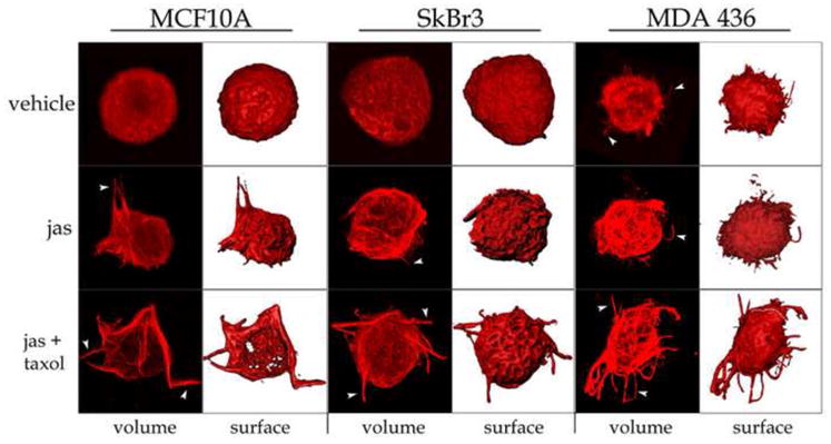
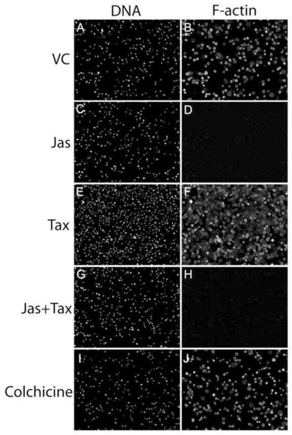
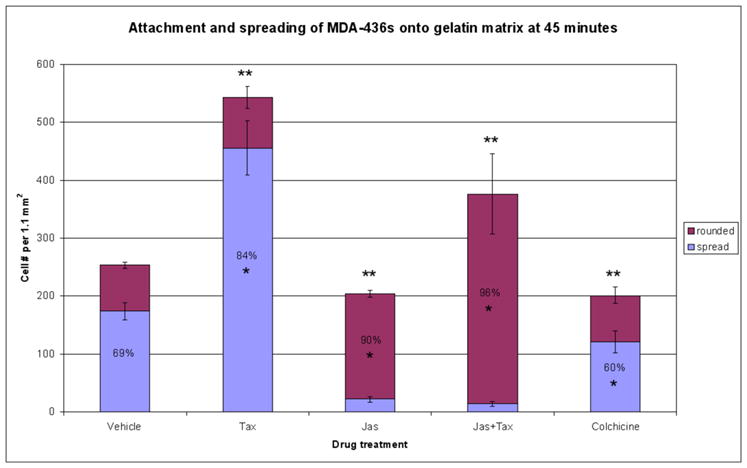
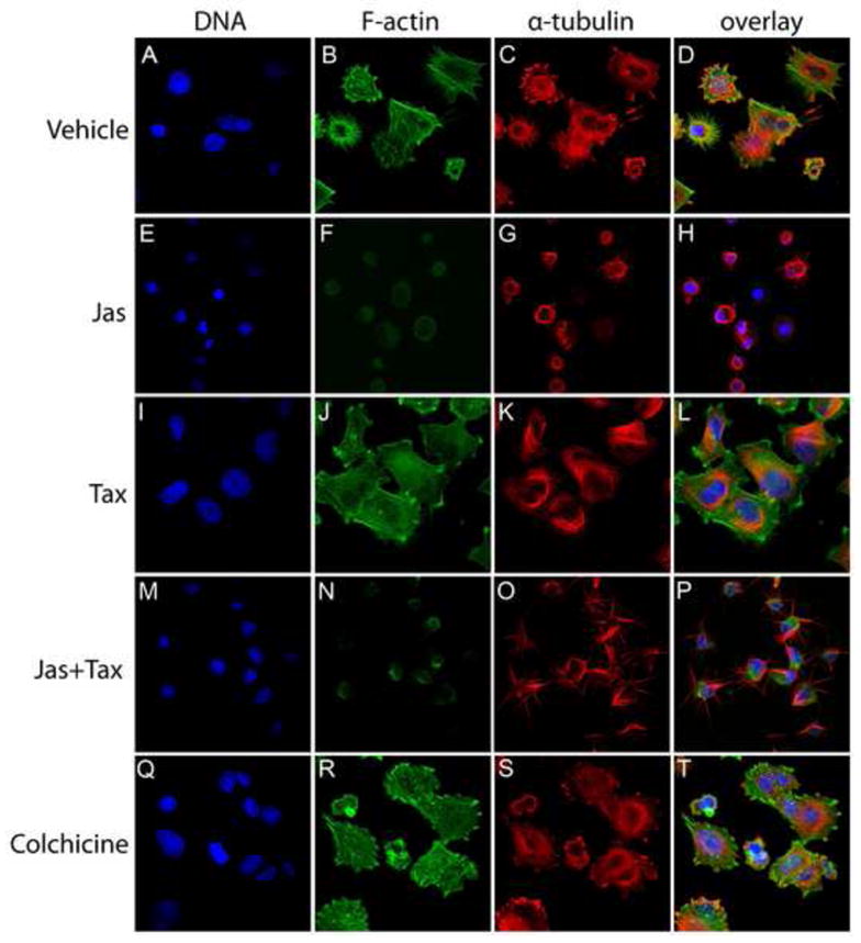
In the clinical treatment of breast cancer, antimitotic cytotoxic agents are one of the most commonly employed chemotherapies, owing largely to their antiproliferative effects on the growth and survival of adherent cells in studies that model primary tumor growth. Importantly, the manner in which these chemotherapeutics impact the metastatic process remains unclear. Furthermore, since dissemination of tumor cells through the systemic circulation and lymphatics necessitates periods of detached survival, it is equally important to consider how circulating tumor cells respond to such compounds. To address this question, we exposed both nontumorigenic and tumor-derived epithelial cell lines to two antitumor compounds, jasplakinolide and paclitaxel (Taxol), in a series of attached and detached states. We report here that jasplakinolide promoted the extension of microtubule-based projections and microtentacle protrusions in adherent and suspended cells, respectively. These protrusions were specifically enriched by upregulation of a stable post-translationally modified form of alpha-tubulin, and this occurred prior to, and independently of any reductions in cellular viability. Microtubule stabilization with Taxol significantly enhanced these effects. Additionally, Taxol promoted the attachment and spreading of suspended tumor cell populations on extracellular matrix. While the antiproliferative effects of these compounds are well recognized and clinically valuable, our findings that microfilament and microtubule binding chemotherapeutics rapidly increase the mechanisms that promote endothelial adhesion of circulating tumor cells warrant caution to avoid inadvertently enhancing metastatic potential, while targeting cell division.


Figures

Figure 1
Figure 1
Filamentous-actin deterioration and MT-stabilization induce α-tubulin projections. To assess the effects of F-actin disruption on the presence and absence of stabilized tubulin, cells were treated with 0.15% DMSO (vehicle; A–D), 1μg/mL Taxol (tax; E–H), 500nM jasplakinolide (jas; I–L), and a combination of Taxol and jas (M-P). Cells were stained for actin with phalloidin (red), antibodies to α-tubulin (green), and Hoescht 33342 (blue) nuclear dye. Taxol reinforced microtubules and promoted the formation of MT bundles and astral bodies (E) that were contained by the microfilament cytoskeleton (F). Jas treatment collapsed F-actin (J), resulting in a loss of regular cell morphology and polymerization of thin microtubule extensions beyond cellular borders (L). Taxol augmented this effect when combined with jas (P). Inset overlay panels (2X mag) reveal the absence of F-actin in tubulin extensions (Scale bar is 10 microns).
Figure 2
Figure 2
Glu-tubulin is enriched in MT-protrusions and upregulated in response to chemotherapeutic agents. Confocal microscopy of cells treated with 0.15% DMSO (vehicle; AC), 1μg/mL Taxol (tax; D–F), 500nM jasplakinolide (jas; G–I), and a combination of jas and Taxol (J–L). Relative to vehicle control cells (A) Taxol increased the abundance of glu-tubulin in all cells examined and caused de novo formation of glu-tubulin-enriched microtubules in the perinuclear region (D). Glu-tubulin was restricted to the cell body and small F-actin membrane spikes were apparent (F, inset). Jas disrupted F-actin (H) and increased glu-tubulin levels and protrusions from the cell body (G). These extensions were devoid of F-actin with particulate glu-tubulin staining (I). Taxol induced enrichment of glu-tubulin in the cellular periphery, and robust membrane protrusions (J). Overlay images confirm the absence of F-actin in protrusions (L, inset panels: 2x mag). (Scale bar is 10 microns).
Figure 3
Figure 3
Jasplakinolide upregulates glu-tubulin. Cells were treated with jas for 1, 3, and 6 hrs, lysed, immuoblotted, and probed for glu-tubulin. MCF-10As showed an increase at out to 3 hrs, and declined to vehicle control (Veh CTL = 0.15% DMSO) levels by 6 hrs. SkBr3 and MDA-436 cells both exhibited significant accumulation of glu-tubulin by 3 hrs, and sustained elevated levels to 6 hrs. α-Tubulin levels indicate even loading of protein samples. Multiple independent lysate sets revealed a consistent trend of increased glu-tubulin (n=3, additional blots not shown).
Figure 4
Figure 4
Chemotherapeutic agents enhance microtentacles in mammary epithelial cells. At least 100 live cells were blindly scored for the presence of microtentacle protrusions, in duplicate over a minimum of three independent trials (n ≥ 6). One hour treatment with 500nM jas increased McTN frequency in nontumorigenic MCF-10As (A), while LA made no significant difference. Both jas and LA induced dramatic increases in McTN score for MDA-436 (B) and SkBr3 (C) breast tumor lines. Taxol augmented these increases all cases, in combination with jas or LA (A–C). Mean values are plotted as percentage of total cells counted + standard error is represented. Significant cell clustering occurred in MCF-10As after suspension in LA + Taxol, so some trials were unable to be quantitated accurately, leading to an n=3 for this specific treatment, rather than an n=6 (*). Carats (^) indicate trials that were significantly increased over 0.15% DMSO vehicle control (Student’s t test, p < 0.01).
Figure 5
Figure 5
Chemotherapeutic agents stimulate MT polymerization and McTN formation. MCF-10A, SkBr3 and MDA-436 cells were treated with 0.15% DMSO vehicle control (top row), 500nM jas (middle row), or a combination of jas and Taxol (bottom row, 1μg/mL) for 1 hour. Cells were suspended for 15 minutes, fixed, and gently spun onto glass coverslips. Cells were then labeled with an antibody against α-tubulin and analyzed by confocal microscopy. Three-dimensional volumes are presented (volume) as well as surface renders to indicate changes in cellular architecture (surface). Treatment with jas or jas+Taxol increased the extension of microtubules beyond the cell periphery in microtentacle protrusions (arrowheads).
Figure 6
Figure 6
Jasplakinolide does not incur cytotoxicity or apoptosis by 6 hrs. (A) MCF-10A, SkBr3, and MDA-436 populations were assayed by colorimetric detection of XTT tetrazolium reduction for alterations in viability after 6 hrs of incubation with 500nM jas. Each of the cell lines tested were viable at the experimental concentration of 500nM, remaining above a value of 1.00, relative to 0.15% DMSO control treated cells. This trend was observed for concentrations up to 10μM (inset, blue line denotes relative viability of 1.00). Bars represent mean + S.E.M for samples from three independent trials.. (B) Individual cells were tested for their ability to exclude propidium iodide (PI). Arrows specify McTN protrusions visible via fluorescent labeling with wheat germ agglutinin (WGA) (top row). PI stained nuclei of cells with compromised membrane integrity, indicated by arrowheads (bottom row). (C) Western blot analysis reveals no significant cleavage of ADP-ribose polymerase (ΔPARP) by 3 hrs in MCF-10As, and by 6 hrs of 500nM jas exposure in the tumor lines examined. TRAIL positively induced PARP cleavage (far right lane). SkBr3 cells exhibit decreased levels of β-actin at each time-point, and MCF-10As appear to upregulate β-actin over the experimental time course, while expression levels were unaltered in MDA-436s. α-tubulin levels indicate even loading of protein samples.
Figure 7
Figure 7
Taxol promotes tumor cell-ECM binding. Equivalent concentrations of MDA-436 cells were plated over gelatin-coated coverslips in serum-free media supplemented with 500nMjas (C and D), 1.17μMTaxol (E and F), jas + Taxol (G and H) or 125μM colchicine (I and J). After 45 minutes, unattached cells were removed by gentle aspiration and washing, and attached cells were fixed and labeled with a nuclear dye (A, C, E, G, I) and a fluorescent phalloidin conjugate (B, D, F, H, J). Images were captured at 10X magnification, revealing differences in total numbers of attached cells.
Figure 8
Figure 8
Taxol significantly enhances tumor cell adhesion and spreading. Five random fields captured at 10X magnification were collected for each drug treatment. Cell nuclei were counted to quantify the number of cells attached within an area of 1.1mm2. Within this area, the fraction of attached cells that remained rounded was also determined via a phalloidin stain, yielding percentages of cell spreading in the presence of each drug. Total cell attachment was inhibited by both jas and colchicine. Taxol was sufficient to significantly enhance adhesion, and restored this effect in jas-treated cells (** denotes significant change in total cell adhesion relative to vehicle control, p<0.001, n=5). Taxol alone increased the percentage of cells that assumed a spread morphology by 45 minutes, while treatments including jas or colchicine impeded cell spreading (* denotes significant percentage change relative to vehicle control, p<0.003, n=5). Percentage labels indicate majority values for each category (i.e. over 50% rounded or spread).
Figure 9
Figure 9
Taxol-treated tumor cells remain flattened and firmly attached beyond 3 hours. Cells were plated over a gelatin matrix in the presence of drug and allowed to settle for 220 minutes. Cells were then fixed and stained with Hoescht 33342 (DNA), phalloidin (F-actin), and an antibody to α-tubulin and analyzed with a laser scanning confocal microscope. Under vehicle control conditions, cell morphology was highly variable (A–D). F-actin was disrupted in jas-treated cells, and MT-rich projections could be seen extending from the cell body (E–H). Co-treatment with Taxol induced a specific enrichment of α-tubulin in the protrusions, relative to the cell body (M–P). Taxol alone stimulated a high percentage of the population to undergo extensive cell spreading (I–L), and some spreading was observed in the colchicine treatment, as F-actin was not perturbed in these cells (Q–T).

References

    1. Mehlen P, Puisieux A. Metastasis: a question of life or death. Nat Rev Cancer. 2006;6:449–58. - PubMed
    1. Chambers AF, Groom AC, MacDonald IC. Dissemination and growth of cancer cells in metastatic sites. Nat Rev Cancer. 2002;2:563–72. - PubMed
    1. Calderwood DA, Shattil SJ, Ginsberg MH. Integrins and actin filaments: reciprocal regulation of cell adhesion and signaling. J Biol Chem. 2000;275:22607–10. - PubMed
    1. Gupton SL, Gertler FB. Filopodia: the fingers that do the walking. Sci STKE. 2007:re5. - PubMed
    1. Weber K, Lazarides E, Goldman RD, Vogel A, Pollack R. Localization and distribution of actin fibers in normal transformed and revertant cells. Cold Spring Harb Symp Quant Biol. 1975;39(Pt 1):363–9. - PubMed