. 2010 Feb;25(2):178-89.
doi: 10.1359/jbmr.090730.

Parathyroid hormone (PTH)-induced bone gain is blunted in SOST overexpressing and deficient mice

Affiliations

Parathyroid hormone (PTH)-induced bone gain is blunted in SOST overexpressing and deficient mice

Ina Kramer et al. J Bone Miner Res. 2010 Feb.

Abstract

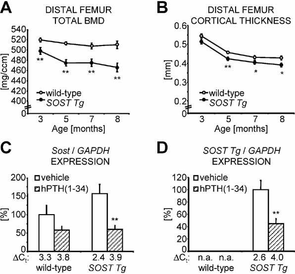
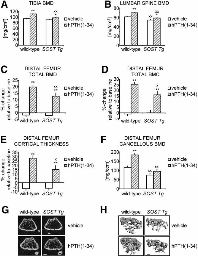
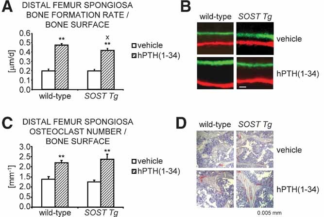
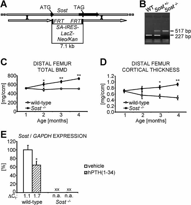
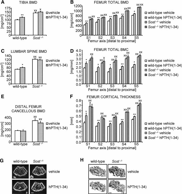
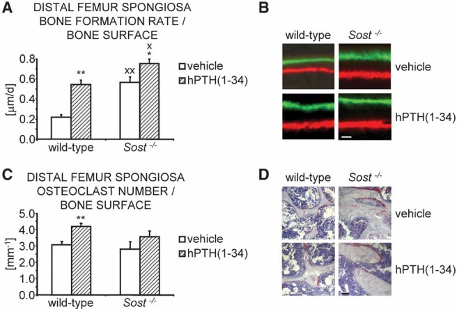
Intermittent parathyroid hormone (PTH) treatment is a potent bone anabolic principle that suppresses expression of the bone formation inhibitor Sost. We addressed the relevance of Sost suppression for PTH-induced bone anabolism in vivo using mice with altered Sost gene dosage. Six-month-old Sost overexpressing and 2-month-old Sost deficient male mice and their wild-type littermates were subjected to daily injections of 100 microg/kg PTH(1-34) or vehicle for a 2-month period. A follow-up study was performed in Sost deficient mice using 40 and 80 microg/kg PTH(1-34). Animals were sacrificed 4 hours after the final PTH administration and Sost expression in long bone diaphyses was determined by qPCR. Bone changes were analyzed in vivo in the distal femur metaphysis by pQCT and ex vivo in the tibia and lumbar spine by DXA. Detailed ex vivo analyses of the femur were performed by pQCT, microCT, and histomorphometry. Overexpression of Sost resulted in osteopenia and Sost deletion in high bone mass. As shown before, PTH suppressed Sost in wild-type mice. PTH treatment induced substantial increases in bone mineral density, content, and cortical thickness and in aging wild-type mice also led to cancellous bone gain owing to amplified bone formation rates. PTH-induced bone gain was blunted at all doses and skeletal sites in Sost overexpressing and deficient mice owing to attenuated bone formation rates, whereas bone resorption was not different from that in PTH-treated wild-type controls. These data suggest that suppression of the bone formation inhibitor Sost by intermittent PTH treatment contributes to PTH bone anabolism.


Figures

Fig. 1
Fig. 1
SOST overexpression results in osteopenia, and SOST is suppressed by PTH. Cross-sectional total BMD (A) and cortical thickness (B) in the distal femur metaphysis were evaluated by monthly in vivo pQCT measurements of skeletally mature to aging wild-type (white data points) and SOST Tg (filled data points) littermate male mice. Relative expression of endogenous mouse Sost (C) and transgenic human SOST (D) in cortical bone of the tibia diaphysis normalized to GAPDH with the corresponding ΔCt values indicated below the bars was determined by qPCR 4 hours following the final PTH (hatched bars) or vehicle (white bars) injection after 2 months of intermittent treatment with 100 µg/kg hPTH(1–34). Data represent means ± SEM. n.a. = not applicable. *p < .05, **p < .01; unpaired Student's t tests for SOST Tg versus wild-type (A, B) or PTH- versus vehicle-treated mice of the same genotype (C, D).
Fig. 2
Fig. 2
PTH-induced bone gain is blunted in SOST Tg mice. Following 2 months of high-dose intermittent PTH treatment, overall BMD in the tibia (A) and the lumbar vertebrae L1–4 (B) was determined in wild-type and SOST Tg mice by ex vivo DXA measurements. Percentage changes relative to baseline were calculated for cross-sectional total BMD (C), BMC (D), and cortical thickness (E) in the distal femur metaphysis, as measured by in vivo pQCT. Cancellous BMD in the distal femur metaphysis (F) was evaluated ex vivo by µCT, as illustrated by the 2D (G) and 3D µCT reconstruction images (H). Data represent means ± SEM for vehicle- (white bars) and PTH-treated (hatched bars) animals. Scale bars: (G) 0.5 mm; (H) 0.25 mm.*,Xp < .05; **,XXp < .01; unpaired Student's t tests for PTH- versus vehicle-treated mice of the same genotype (*, **) or SOST Tg versus wild-type mice of the same treatment group (X, XX).
Fig. 3
Fig. 3
PTH-induced increase in bone formation rate is attenuated in SOST Tg mice. (A) Bone formation rates were determined by dynamic histomorphometry in the cancellous compartment of the distal femur metaphysis of vehicle- (white bars) and PTH-treated (hatched bars) wild-type and SOST Tg mice. (B) Representative images of fluorochrome-marker-labeled trabecular bone surfaces. (C) Osteoclast number was evaluated on TRAP-stained femoral sections, as illustrated by representative images of the cancellous compartment of the distal femur metaphysis (D). Scale bars: (B) 0.5 µm; (D) 5 µm. Data represent means ± SEM. Xp < .05; **p < 0.01; unpaired Student's t tests for PTH- versus vehicle-treated mice of the same genotype (**) or SOST Tg versus wild-type mice of the same treatment group (X).
Fig. 4
Fig. 4
Sost loss-of-function results in a progressive high bone mass phenotype. (A) Genomic targeting scheme depicting the recombination strategy for generating the targeted Sost KO allele in which a 7.1-kb LacZ-neomycin(Neo)/kanamycin(Kan) resistance selection cassette replaces most of the Sost coding region, indicated by the ATG translation initiation and the TAG stop codon. Homology arms present in the targeting construct are represented by gray arrows; the 2 exons of the Sost gene are shown as black arrows. Flipase recognition target (FRT) sites for optional removal of the selection cassette are depicted as black boxes flanking the selection cassette. IRES = internal ribosome entry site; SA = splice acceptor site. (B) Genomic DNA was extracted from tail tips and used for genotyping of offspring obtained from heterozygous (Sost+/−) Sost KO breeding pairs to generate wild-type (WT), heterozygous, and homozygous (Sost−/−) Sost KO mice. Cross-sectional total BMD (C) and cortical thickness (D) in the distal femur metaphysis of wild-type (white data points) and Sost−/− (filled data points) littermate male mice were evaluated at monthly intervals starting at 1 month of age until skeletal maturity. (E) 4 hours after the final PTH or vehicle application following 2 months of intermittent treatment with 100 µg/kg hPTH(1–34), the relative expression of Sost normalized to GAPDH, with the corresponding ΔCt values indicated below the bars, was determined by qPCR in cortical bone of the femur diaphysis. Data represent means ± SEM. n.a. = not applicable. *p < .05, **p < .01; unpaired Student's t tests for Sost−/− versus wild-type mice (C, D) or PTH- versus vehicle-treated mice of the same genotype (E). XXp < .01; unpaired Student's t tests for Sost−/− versus wild-type mice of the same treatment group (E).
Fig. 5
Fig. 5
PTH-induced bone gain is blunted in Sost−/− mice. Overall BMD in the tibia (A) and the lumbar vertebrae L1–4 (C) was determined in wild-type and Sost KO male mice ex vivo by DXA analyses following 2 months of high-dose intermittent PTH treatment. Femoral cross-sectional total BMD (B), BMC (D), and cortical thickness (F) were assessed by ex vivo pQCT measurements of five consecutive slices S1 to S5 distributed along the femoral axis at equal distances from the distal metaphysis (S1) to the proximal end of the diaphysis (S5). Cancellous BMD in the distal femur metaphysis (E) was evaluated ex vivo by µCT, as illustrated by the 2D (G) and 3D µCT reconstruction images (H). Scale bars: (G) 0.5 mm; (H) 0.25 mm. Data represent means ± SEM for vehicle- (white bars) and PTH-treated (hatched bars) animals. *,Xp < .05; **,XXp < .01; unpaired Student's t tests for PTH- versus vehicle-treated mice of the same genotype (*, **) or Sost−/− versus wild-type mice of the same treatment group (X, XX).
Fig. 6
Fig. 6
PTH-induced increase in bone formation rate is blunted in Sost−/− mutant mice. (A) Bone formation rates were determined by dynamic histomorphometry in the cancellous compartment of the distal femur metaphysis of vehicle- (white bars) and PTH-treated (hatched bars) wild-type and Sost−/− mice. (B) Representative images of fluorochrome-marker-labeled trabecular bone surfaces. (C) Osteoclast number was evaluated on TRAP-stained femoral sections, as illustrated by representative images of the cancellous compartment of the distal femur metaphysis (D). Scale bars: (B) 0.5 µm; (D) 5 µm. Data represent means ± SEM. *,Xp < .05; **,XXp < .01; unpaired Student's t tests for PTH- versus vehicle-treated mice of the same genotype (*, **) or Sost−/− versus wild-type mice of the same treatment group (X, XX).
Fig. 7
Fig. 7
PTH-induced bone gain is blunted in Sost−/− mice irrespective of the applied PTH dose. (A) Cross-sectional total BMD in the proximal tibia metaphysis was determined by in vivo pQCT analyses in wild-type and Sost−/− male mice after 2 months of intermittent treatment with daily injections of vehicle (white bars) or 40 (fine hatched bars) or 80 µg/kg hPTH(1–34) (bold hatched bars). (B) Cancellous BMD was evaluated in the distal femur metaphysis by ex vivo µCT. (C) Femoral cross-sectional total BMD, total BMC (D), and cortical thickness (E) were evaluated by ex vivo pQCT analyses of five consecutive slices S1 to S5 distributed along the femoral axis at equal distances from the distal metaphysis (S1) to the proximal end of the diaphysis (S5). Data represent means ± SEM. *,Xp < .05; **,XXp < .01; unpaired Student's t tests for PTH- versus vehicle-treated mice of the same genotype (*, **) or Sost−/− versus wild-type mice of the same treatment group (X, XX).

Comment in

References

    1. Bonnick SL. Osteoporosis in men and women. Clin Cornerstone. 2006;8:28–39. - PubMed
    1. Hodsman AB, Bauer DC, Dempster DW, et al. Parathyroid hormone and teriparatide for the treatment of osteoporosis: a review of the evidence and suggested guidelines for its use. Endocr Rev. 2005;26:688–703. - PubMed
    1. Khosla S, Westendorf JJ, Oursler MJ. Building bone to reverse osteoporosis and repair fractures. J Clin Invest. 2008;118:421–428. - PMC - PubMed
    1. Juppner H, Abou-Samra AB, Freeman M, et al. A G protein-linked receptor for parathyroid hormone and parathyroid hormone-related peptide. Science. 1991;254:1024–1026. - PubMed
    1. Nishida S, Yamaguchi A, Tanizawa T, et al. Increased bone formation by intermittent parathyroid hormone administration is due to the stimulation of proliferation and differentiation of osteoprogenitor cells in bone marrow. Bone. 1994;15:717–723. - PubMed

Publication types

MeSH terms