Review
. 2009 Nov;9(5):493-506.
doi: 10.2174/187152609789105687.

The Seattle Structural Genomics Center for Infectious Disease (SSGCID)

Affiliations
Review

The Seattle Structural Genomics Center for Infectious Disease (SSGCID)

P J Myler et al. Infect Disord Drug Targets. 2009 Nov.

Abstract

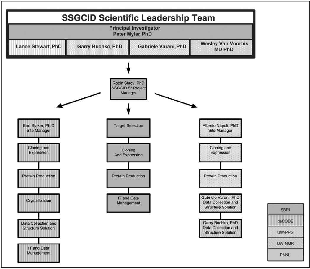
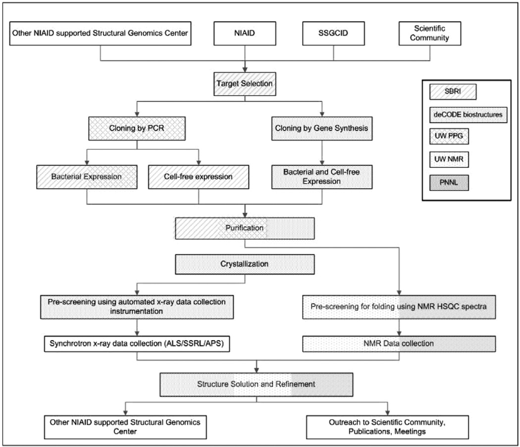
The NIAID-funded Seattle Structural Genomics Center for Infectious Disease (SSGCID) is a consortium established to apply structural genomics approaches to potential drug targets from NIAID priority organisms for biodefense and emerging and re-emerging diseases. The mission of the SSGCID is to determine approximately 400 protein structures over the next five years. In order to maximize biomedical impact, ligand-based drug-lead discovery campaigns will be pursued for a small number of high-impact targets. Here we review the center's target selection processes, which include pro-active engagement of the infectious disease research and drug therapy communities to identify drug targets, essential enzymes, virulence factors and vaccine candidates of biomedical relevance to combat infectious diseases. This is followed by a brief overview of the SSGCID structure determination pipeline and ligand screening methodology. Finally, specifics of our resources available to the scientific community are presented. Physical materials and data produced by SSGCID will be made available to the scientific community, with the aim that they will provide essential groundwork benefiting future research and drug discovery.


Figures

Fig. 1
Fig. 1. SSGCID Management and Organization
SBRI: Seattle Biomedical Research Institute, deCODE: deCODE biostructures, UW-PPG: University of Washington Protein Production Group, UW-NMR: University of Washington NMR Group, PNNL: Pacific Northwest National Laboratory.
Fig. 2
Fig. 2. SSGCID Structure Determination Pipeline
Inset box shows code for sites performing Target Selection, Cloning & Expression Screening, Protein Production, Crystallization, and Data Collection & Structure Solution.
Fig. 3
Fig. 3. Selected protein structures from SSGCID
Panel I. The solution structure for BolA-like protein PFE0790c from P. falciparum (PDB ID: 2KDN). The ribbon image on the left represents the ensemble of the final 20 NMR structures superimposed on the average structure, while the right cartoon represents the structure closest to the average structure with the three α-helices and three β-strands labeled. For clarity, the unstructured, N-terminal 22-residue tag has been removed from both structures. Color scheme: Helices = red, β-strands = cyan, loops and turns = grey. Panel II. Ribbon drawing of the RNA methyltransferase BupsA.00072.a from B. pseudomallei (3E5Y). The asymmetric unit contains a dimer of two molecules, which is the biological unit. On the left dimer, the thread of the knot can be seen as the orange-red section passing through the yellow-green section. These are roughly residues 80–120 of the 156 amino acid protein. Panel III. Ligand-bound structures of BupsA.00114.a, phosphoglycerate mutase from B. pseudomallei (3EZN). The reaction catalyzed by this enzyme is shown in the top panel. Close-ups of the active site of the phosphoglycerate mutase reveal the 3PG substrate and a transition-state intermediate as a covalently-bound phosphate (left panel), which can be mimicked by vanadate + glycerol (center panel). The final product, 2,3-BPG is shown in the right panel. Panel IV. Fragment-bound Structures from BupsA.00027.a, a glutaryl-CoA dehydrogenase from B. pseudomallei (3D6B). The left panel contains a ribbon diagram of BupsA.00027.a colored by secondary structure, showing α-helices (red), and β-sheets (yellow). Three different fragments (cyan, pink and purple) are superimposed/bound in the active-site. The right panel shows a close-up up of active-site binding pocket, showing the same three fragments. Panel V. Hexameric structure of inorganic pyrophosphatase, RiprA.00023.a, from Rickettsia prowazekii (3D53). Each 20 kDa monomer is colored differently (magenta, gray, green, yellow, pink, peach). Panel VI. Endogenous co-factor (NAD) bound to the glyceraldehyde-3-phosphate dehydrogenase, BrabA.00052.a, from B. melitenesis (3DOC) The NAD molecule is shown as a space-filling model within the ribbon diagram of the protein.

References

    1. Wishart DS, Knox C, Guo AC, Shrivastava S, Hassanali M, Stothard P, Chang Z, Woolsey J. Nucleic Acids Res. 2006;34(Database issue):D668–D672. - PMC - PubMed
    1. Krogh A, Larsson B, Von H, Sonnhammer EL. J. Mol. Biol. 2001;305(3):567–580. - PubMed
    1. Edwards TE, Ferre-D'Amare AR. Structure. 2006;14(9):1459–1468. - PubMed
    1. Aslanidis C, De Jong PJ. Nucleic. Acids Res. 1990;18:6069–6074. - PMC - PubMed
    1. Alexandrov A, Vignali M, LaCount DJ, Quartley E, de Vries C, De Rosa D, Babulski J, Mitchell SF, Schoenfeld LW, Fields S, Hol WG, Dumont ME, Phizicky EM, Grayhack EJ. Mol. Cell. Proteomics. 2004;3(9):934–938. - PubMed

Publication types

LinkOut - more resources