

Review
doi: 10.1186/1475-2875-8-153.
Computer vision for microscopy diagnosis of malaria
Affiliations
- PMID: 19594927
- PMCID: PMC2719653
- DOI: 10.1186/1475-2875-8-153
Item in Clipboard
Review
Computer vision for microscopy diagnosis of malaria
Malar J.
.
Abstract
This paper reviews computer vision and image analysis studies aiming at automated diagnosis or screening of malaria infection in microscope images of thin blood film smears. Existing works interpret the diagnosis problem differently or propose partial solutions to the problem. A critique of these works is furnished. In addition, a general pattern recognition framework to perform diagnosis, which includes image acquisition, pre-processing, segmentation, and pattern classification components, is described. The open problems are addressed and a perspective of the future work for realization of automated microscopy diagnosis of malaria is provided.
Figures
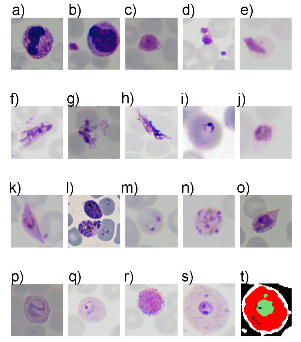
Examples of stained objects. (a, b) white blood cells, (c, d) platelets, (e)-(h) artefacts, (i)-(l) P. falciparum ring, trophozoite, gametocyte, schizont, (m, n) P. malariae ring and schizont (o, p) P. ovale and P. vivax trophozoites, (q, r) P. vivax ring and gametocyte, (s) P. vivax ring, (t) extracted stained pixel group. (S, green region(s)) and the stained object (Sb, red region including the green one).
Stained object classes: in a Giemsa-stained blood film an observed stained object can be a parasite from one of the four species of Plasmodium or a regular blood component such as white blood cell, platelet. Artefact class represents bacteria, spores, crystallized stain chemicals, particles due to dirt, RBC anomalies (e.g. Howell-Jolly bodies, iron deficiency, reticulocytes), and other peripheral blood parasites.
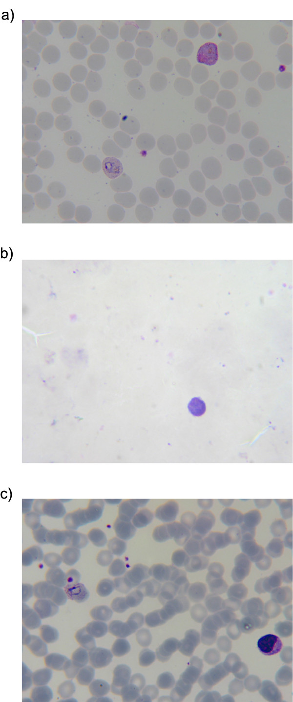
Examples of Giemsa-stained (a) thin and (b) thick blood film smear images, (c) a concentrated (thick) field of a thin blood film smear.
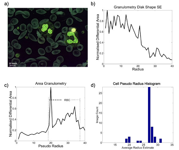
Size granulometry vs. area granulometry. (a) negative image of the grey level sickle cell image, (b) granulometry using disk shaped structuring elements, (c) area granulometry, (d) area granulometry based cell size estimation varies in different fields of a thin film although the magnification is constant.
Similar articles
-
[Advances in automatic detection technology for images of thin blood film of malaria parasite].Zhongguo Xue Xi Chong Bing Fang Zhi Za Zhi. 2017 May 5;29(3):388-392. doi: 10.16250/j.32.1374.2017015. Zhongguo Xue Xi Chong Bing Fang Zhi Za Zhi. 2017. PMID: 29469543 Review. Chinese.
-
Microscopic malaria parasitemia diagnosis and grading on benchmark datasets.Microsc Res Tech. 2018 Sep;81(9):1042-1058. doi: 10.1002/jemt.23071. Epub 2018 Sep 12. Microsc Res Tech. 2018. PMID: 30207623 Review.
-
Web-Enabled Distributed Health-Care Framework for Automated Malaria Parasite Classification: an E-Health Approach.J Med Syst. 2017 Oct 26;41(12):192. doi: 10.1007/s10916-017-0834-0. J Med Syst. 2017. PMID: 29075939
-
Image processing and 2-D morphometry.Curr Protoc Cytom. 2001 May;Chapter 10:Unit 10.11. doi: 10.1002/0471142956.cy1011s13. Curr Protoc Cytom. 2001. PMID: 18770675
-
Application of principal component analysis to multispectral-multimodal optical image analysis for malaria diagnostics.Malar J. 2014 Dec 11;13:485. doi: 10.1186/1475-2875-13-485. Malar J. 2014. PMID: 25495235 Free PMC article.
Cited by
-
Automated wide-field malaria parasite infection detection using Fourier ptychography on stain-free thin-smears.Biomed Opt Express. 2022 Jun 15;13(7):3904-3921. doi: 10.1364/BOE.448099. eCollection 2022 Jul 1. Biomed Opt Express. 2022. PMID: 35991917 Free PMC article.
-
Exploring the Impact of Batch Size on Deep Learning Artificial Intelligence Models for Malaria Detection.Cureus. 2024 May 13;16(5):e60224. doi: 10.7759/cureus.60224. eCollection 2024 May. Cureus. 2024. PMID: 38868293 Free PMC article.
-
Deep Learning Based Automatic Malaria Parasite Detection from Blood Smear and its Smartphone Based Application.Diagnostics (Basel). 2020 May 20;10(5):329. doi: 10.3390/diagnostics10050329. Diagnostics (Basel). 2020. PMID: 32443868 Free PMC article.
-
PERCEPTION OF HEALTH CARE WORKERS ON ARTIFICIAL INTELLIGENCE BASED MALARIA DIAGNOSIS IN SOUTHWESTERN NIGERIA.Ann Ib Postgrad Med. 2024 Dec 31;22(3):16-21. Ann Ib Postgrad Med. 2024. PMID: 40385718 Free PMC article.
-
Computer Vision Malaria Diagnostic Systems-Progress and Prospects.Front Public Health. 2017 Aug 21;5:219. doi: 10.3389/fpubh.2017.00219. eCollection 2017. Front Public Health. 2017. PMID: 28879175 Free PMC article. Review.
References
-
- Korenromp E, Miller J, Nahlen B, Wardlaw T, Young M. Tech rep. World Health Organization, Geneva; 2005. World Malaria Report 2005.
-
- WHO . Basic malaria microscopy Part I Learner's Guide. World Health Organization; 1991.
-
- Coleman RE, Maneechai N, Rachaphaew N, Kumpitak C, Miller R, Soyseng V, Thimasarn K, Sattabongkot J. Comparison of field and expert laboratory microscopy for active surveillance for asymptomatic Plasmodium falciparum and Plasmodium vivax in Western Thailand. Am J Trop Med Hyg. 2002;67:141–144. - PubMed
Publication types
MeSH terms
LinkOut - more resources
Full Text Sources
Other Literature Sources
Medical
