

Preparation and biological properties of a novel composite scaffold of nano-hydroxyapatite/chitosan/carboxymethyl cellulose for bone tissue engineering
- PMID: 19594953
- PMCID: PMC2720940
- DOI: 10.1186/1423-0127-16-65
Preparation and biological properties of a novel composite scaffold of nano-hydroxyapatite/chitosan/carboxymethyl cellulose for bone tissue engineering
Abstract
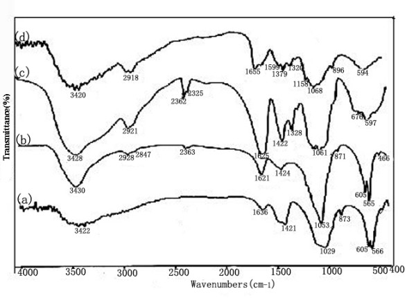
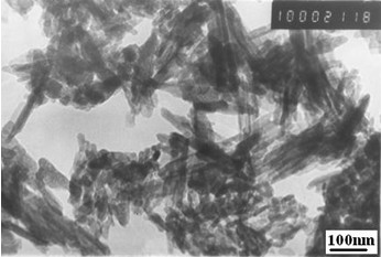
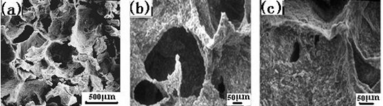
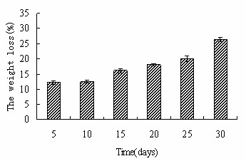
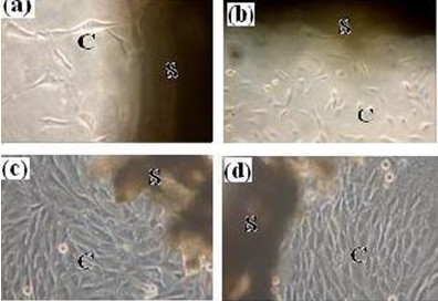
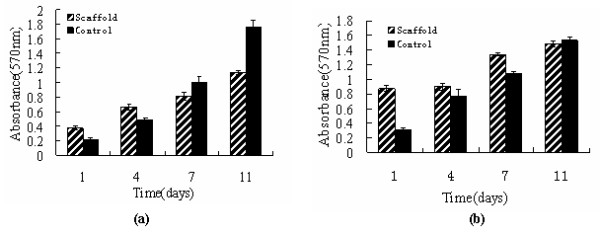
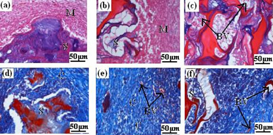
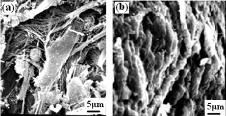
In this study, we report the physico-chemical and biological properties of a novel biodegradable composite scaffold made of nano-hydroxyapatite and natural derived polymers of chitosan and carboxymethyl cellulose, namely, n-HA/CS/CMC, which was prepared by freeze-drying method. The physico-chemical properties of n-HA/CS/CMC scaffold were tested by infrared absorption spectra (IR), transmission electron microscope(TEM), scanning electron microscope(SEM), universal material testing machine and phosphate buffer solution (PBS) soaking experiment. Besides, the biological properties were evaluated by MG63 cells and Mesenchymal stem cells (MSCs) culture experiment in vitro and a short period implantation study in vivo. The results show that the composite scaffold is mainly formed through the ionic crossing-linking of the two polyions between CS and CMC, and n-HA is incorporated into the polyelectrolyte matrix of CS-CMC without agglomeration, which endows the scaffold with good physico-chemical properties such as highly interconnected porous structure, high compressive strength and good structural stability and degradation. More important, the results of cells attached, proliferated on the scaffold indicate that the scaffold is non-toxic and has good cell biocompatibility, and the results of implantation experiment in vivo further confirm that the scaffold has good tissue biocompatibility. All the above results suggest that the novel degradable n-HA/CS/CMC composite scaffold has a great potential to be used as bone tissue engineering material.
Figures
References
-
- Hirotaka M, Toshihiro K, Masayuki N, Minoru U. Preparation of bonelike apatite composite for tissue engineering scaffold. Sci Technol Adv Mat. 2005;6:48–53. doi: 10.1016/j.stam.2004.07.003. - DOI
-
- Wang YJ, Yang CR, Chen XF, Zhao NR. Development and Characterization of Novel Biomimetic Composite Scaffolds Based on Bioglass-Collagen-Hyaluronic Acid-Phosphatidylserine for Tissue Engineering Applications. Macromol Mater Eng. 2006;291:254–262. doi: 10.1002/mame.200500381. - DOI
Publication types
MeSH terms
Substances
LinkOut - more resources
Full Text Sources
Other Literature Sources
