. 2009 Oct;30(29):5433-44.
doi: 10.1016/j.biomaterials.2009.06.042.

Mechanosensitivity of fibroblast cell shape and movement to anisotropic substratum topography gradients

Affiliations

Mechanosensitivity of fibroblast cell shape and movement to anisotropic substratum topography gradients

Deok-Ho Kim et al. Biomaterials. 2009 Oct.

Abstract

In this report, we describe using ultraviolet (UV)-assisted capillary force lithography (CFL) to create a model substratum of anisotropic micro- and nanotopographic pattern arrays with variable local density for the analysis of cell-substratum interactions. A single cell adhesion substratum with the constant ridge width (1 microm), and depth (400 nm) and variable groove widths (1-9.1 microm) allowed us to characterize the dependence of cellular responses, including cell shape, orientation, and migration, on the anisotropy and local density of the variable micro- and nanotopographic pattern. We found that fibroblasts adhering to the denser pattern areas aligned and elongated more strongly along the direction of ridges, vs. those on the sparser areas, exhibiting a biphasic dependence of the migration speed on the pattern density. In addition, cells responded to local variations in topography by altering morphology and migrating along the direction of grooves biased by the direction of pattern orientation (short term) and pattern density (long term), suggesting that single cells can sense the topography gradient. Molecular dynamic live cell imaging and immunocytochemical analysis of focal adhesions and actin cytoskeleton suggest that variable substratum topography can result in distinct types of cytoskeleton reorganization. We also demonstrate that fibroblasts cultured as monolayers on the same substratum retain most of the properties displayed by single cells. This result, in addition to demonstrating a more sophisticated method to study aspects of wound healing processes, strongly suggests that even in the presence of adhesive cell-cell interactions, the cues provided by the underlying substratum topography continue to exercise substantial influence on cell behavior. The described experimental platform might not only further our understanding of biomechanical regulation of cell-matrix interactions, but also contribute to bioengineering of devices with the optimally structured design of cell-material interface.


Figures

Figure 1
Figure 1
Fabrication of polymeric ridge pattern arrays with graded spacing. (a) Schematic of the microfabrication process used to generate PUA micro- and nanopattern arrays. Drawings are not to scale. (b) The three-dimensional AFM image of topographic ridge pattern arrays of ∼400 nm height and ∼1 µm width. (c) An SEM image of one-dimensional topographical ridge pattern with graded spacing (∼100 nm increments in groove width between neighboring ridges).
Figure 2
Figure 2
The morphological response of fibroblasts to the local density of topographic pattern arrays with graded spacing. (a) A phase-contrast micrograph of NIH 3T3 fibroblasts shows the differential morphology associated with the density variation of the underlying topographic pattern arrays. (b–c) SEM images of critical point-dried NIH 3T3 fibroblasts plated for 14 hr (b) on variable ridge pattern arrays with graded spacing and (c) on regularly spaced topographic pattern arrays with 1 µm wide ridges and 1 µm wide grooves. The white arrow indicates membrane protrusion extending toward the more densely spaced ridges.
Figure 3
Figure 3
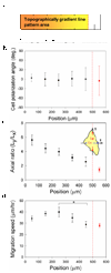
Effects of the local density of ridged pattern arrays on cell shape and migration. (a) A schematic representation of the density gradient of the ridged pattern array, indicating the distance measured from the most densely patterned area (0∼100 µm) to the smooth area (500∼600 µm). The average groove width is 2.6 µm, 4.8 µm, 6.3 µm, 7.5 µm and 8.6 µm in these zones, ranging from the most densely patterned area (0∼100 µm) to the smooth area (500∼600 µm), respectively. (b) The distribution of orientation angles for cells adhered to the substratum with the graded ridge spacing (error bars represent mean ± SD). (c) Morphological changes of fibroblasts as a function of the density variations in the underlying substratum, described by axial ratio of Ly/Lx as a function of position (error bars represent mean ± SEM). (d) A biphasic dependence of cell migration speed on the topographic density (error bars represent mean ± SEM).
Figure 4
Figure 4
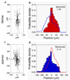
Analysis of cell migration trajectories. Migration trajectories of cells (a) on a densely spaced ridged arrays (positioned between 0 µm and 250 µm; the range of groove width: 1 µm ∼ 6.3 µm; n=27) and (c) on a sparsely spaced ridged arrays (positioned between 250 µm and 500 µm; the range of groove width: 6.3 µm ∼ 9.1 µm; n=55). Probability of a cell trajectory to encounter the zone of a given ridge density on the dense (b) or sparse (d) ridge arrays. Skewness of the probability distributions is shown in each panel (b, d). Distributions indicated in each panel suggest that cell trajectories are biased toward the sparser pattern area for cells on dense ridge arrays (positively skewed distribution in (b)) and towards the denser pattern area for cells on sparse ridge arrays (negatively skewed distribution in (d)).
Figure 5
Figure 5
Representative immunofluorescent micrographs of fibroblasts cultured on the patterned ridged substrata of different local densities. (a) A cell on the most densely spaced ridged arrays (positioned between 0 µm and 100 µm; the range of groove width: 1 µm ∼ 3.8 µm). (b) A cell on the most sparsely spaced ridged arrays (positioned between 400 µm and 500 µm; the range of groove width: 8.1 µm ∼ 9.1 µm). (c–d) Spatial co-localization of the actin cytoskeleton and FAs in the protruding lamelipodium aligned proximal to the individual ridge. (e) Quantitative and correlative analysis of the actin cytoskeleton and FAs in the protruding lamelipodium (error bars represent mean ± SEM).
Figure 6
Figure 6
Effects of substratum topography on adhesion dynamics. (a–b) Time-lapse photographs of FA orientations of NIH 3T3 fibroblast cells transfected with GFP-vinculin cultured on (a) the densely spaced ridged array and (b) the sparsely spaced ridged array. (c–d) Angular histograms showing the distribution of FA orientation dynamically changing over time after exposed to the underlying (c) dense (positioned between 0 µm and 200 µm; the range of groove width: 1 µm ∼ 5.6 µm) and (d) sparse (positioned between 300 µm and 500 µm; the range of groove width: 6.9 µm ∼ 9.1 µm) ridged pattern arrays. The average groove width of the underlying patterns is 3.1 µm in (a) and 8.1 µm in (b).
Figure 7
Figure 7
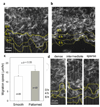
Topography-induced change in collective fibroblast cell migration (a–b) Phase contrast micrographs of collective fibroblast migration on (a) anisotropic micro- and nanopatterns and (b) the flat substratum in an in vitro wound healing model. (c) Quantification of cell migration speed for 12 hr (error bars represent mean ± SEM). (d) Representative time-lapse photographs showing the progression of collective fibroblast migration from dense (the average groove width: 3 µm), intermediate (the average groove width: 6.8 µm), and sparse (the average groove width: 8.7 µm) pattern density areas, respectively. The range of groove width in (a) is 3.8 µm and 6.4 µm.
Figure 8
Figure 8
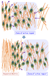
Model of the role of fibroblasts in extracellular matrix remodeling during wound healing. During the wound healing process, fibroblasts are functionally essential cells that migrate toward and into the open wound over the fibrin clot, which results in deposition of collagen, restructuring of ECM and formation of granulation tissue. Migrating fibroblasts also exert tractional forces onto the collagen matrix, which results in its reorganization along the direction of the resulting stress. Our results suggest that fibroblasts can aggregate in zones of intermediate ECM density that might become the zones of active ECM repair, which can shift as cells progressively create denser matrix, following which cells clear these now denser zones and shift to the adjacent zone. This, along with apoptosis, can be an efficient mechanism of cell clearance from the restructured wound zones prior to re-epithelialization.

Similar articles

Cited by

References

    1. Kim DH, Wong PK, Park J, Levchenko A, Sun Y. Microengineered platforms for cell mechanobiology. Annu Rev Biomed Eng. 2009;11:203–233. - PubMed
    1. Dalby MJ, Gadegaard N, Tare R, Andar A, Riehle MO, Herzyk P, et al. The control of human mesenchymal cell differentiation using nanoscale symmetry and disorder. Nat Mater. 2007;6(12):997–1003. - PubMed
    1. Patel S, Kurpinski K, Quigley R, Gao H, Hsiao BS, Poo MM, et al. Bioactive nanofibers: synergistic effects of nanotopography and chemical signaling on cell guidance. Nano Lett. 2007;7(7):2122–2128. - PubMed
    1. Kim DH, Levchenko A, Suh KY. Engineered surface nanotopography for controlling cell-substrate interactions. In: Khademhosseini A JB, Takayama S, Toner M, editors. Micro- and Nanoengineering of the Cell Microenvironment: Technologies and Applications. Boston: Artech House; 2008. pp. 185–208.
    1. Ottani V, Raspanti M, Ruggeri A. Collagen structure and functional implications. Micron. 2001;32(3):251–260. - PubMed

Publication types

Substances