

Nanoparticles evading the reticuloendothelial system: role of the supported bilayer
- PMID: 19595666
- PMCID: PMC2757503
- DOI: 10.1016/j.bbamem.2009.06.022
Nanoparticles evading the reticuloendothelial system: role of the supported bilayer
Abstract
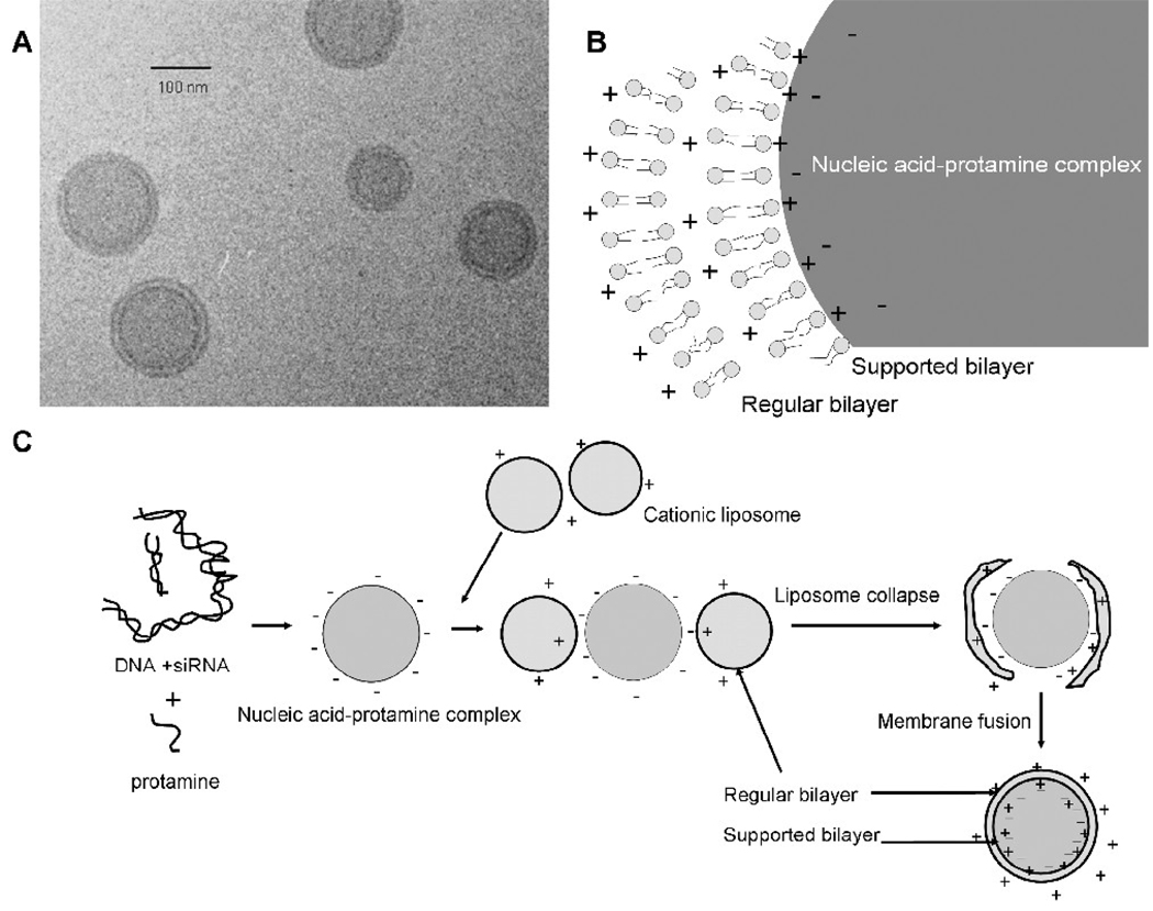
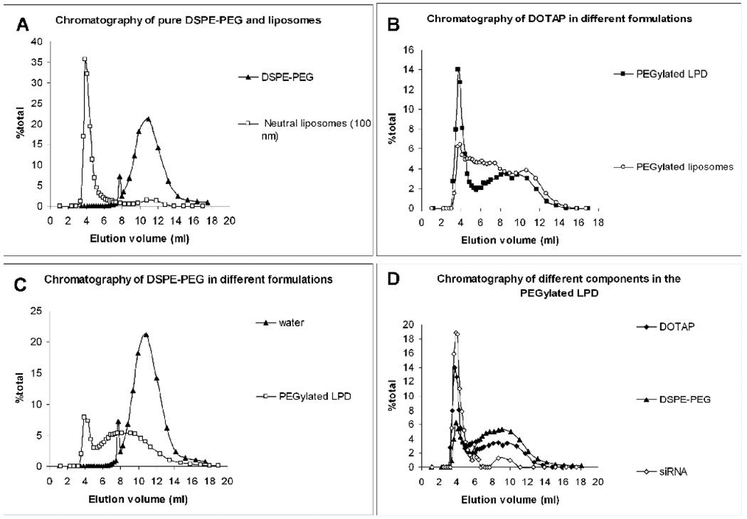
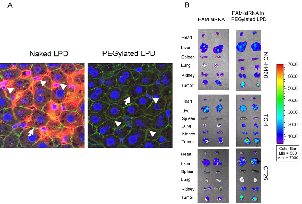
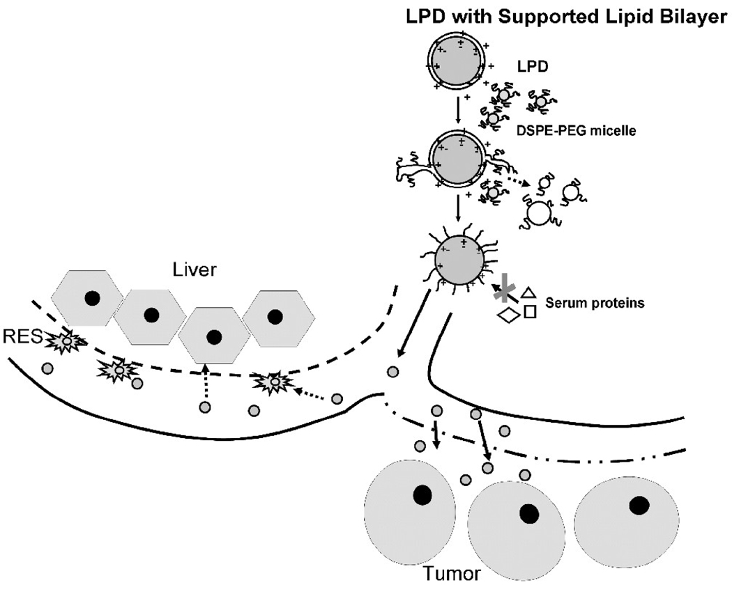
We have previously shown that the PEGylated LPD (liposome-polycation-DNA) nanoparticles were highly efficient in delivering siRNA to the tumor with low liver uptake. Its mechanism of evading the reticuloendothelial system (RES) is reported here. In LPD, nucleic acids were condensed with protamine into a compact core, which was then coated by two cationic lipid bilayers with the inner bilayer stabilized by charge-charge interaction (also called the supported bilayer). Finally, a detergent-like molecule, polyethylene glycol (PEG)-phospholipid is post-inserted into the lipid bilayer to modify the surface of LPD. The dynamic light scattering (DLS) data showed that LPD had improved stability compared to cationic liposomes after incubation with a high concentration of DSPE-PEG(2000), which is known to disrupt the bilayer. LPD prepared with a multivalent cationic lipid, DSGLA, had enhanced stability compared to those containing DOTAP, a monovalent cationic lipid, suggesting that stronger charge-charge interaction in the supported bilayer contributed to a higher stability. Distinct nanoparticle structure was found in the PEGylated LPD by transmission electron microscopy, while the cationic liposomes were transformed into tubular micelles. Size exclusion chromatography data showed that approximately 60% of the total cationic lipids, which were located in the outer bilayer of LPD, were stripped off during the PEGylation; and about 20% of the input DSPE-PEG(2000) was incorporated into the inner bilayer with about 10.6 mol% of DSPE-PEG(2000) presented on the particle surface. This led to complete charge shielding, low liver sinusoidal uptake, and 32.5% injected dose delivered to the NCI-H460 tumor in a xenograft model.
Figures


References
-
- Klibanov AL, Maruyama K, Torchilin VP, Huang L. Amphipathic polyethyleneglycols effectively prolong the circulation time of liposomes. FEBS Lett. 1990;268:235–237. - PubMed
-
- Woodle MC, Lasic DD. Sterically stabilized liposomes. Biochim Biophys Acta. 1992;1113:171–199. - PubMed
-
- Uster PS, Allen TM, Daniel BE, Mendez CJ, Newman MS, Zhu GZ. Insertion of poly(ethylene glycol) derivatized phospholipid into pre-formed liposomes results in prolonged in vivo circulation time. FEBS Lett. 1996;386:243–246. - PubMed
-
- Silvander M, Johnsson M, Edwards K. Effects of PEG-lipids on permeability of phosphatidylcholine/cholesterol liposomes in buffer and in human serum. Chem Phys Lipids. 1998;97:15–26. - PubMed
-
- Li S, Huang L. In vivo gene transfer via intravenous administration of cationic lipid-protamine-DNA (LPD) complexes. Gene Ther. 1997;4:891–900. - PubMed